5FDI | pdb_00005fdi

Crystal structure of Human Carbonic Anhydrase II with the anticonvulsant sulfamide JNJ-26990990 and its S,S-dioxide analog.


Experimental Data Snapshot

  • Method: X-RAY DIFFRACTION
  • Resolution: 1.85 Å
  • R-Value Free: 
    0.193 (Depositor), 0.190 (DCC) 
  • R-Value Work: 
    0.162 (Depositor), 0.158 (DCC) 

Starting Model: experimental
View more details

wwPDB Validation 3D Report Full Report

Validation slider image for 5FDI

Ligand Structure Quality Assessment 


This is version 1.2 of the entry. See complete history

Literature

The anticonvulsant sulfamide JNJ-26990990 and its S,S-dioxide analog strongly inhibit carbonic anhydrases: solution and X-ray crystallographic studies.

Di Fiore, A.De Simone, G.Alterio, V.Riccio, V.Winum, J.Y.Carta, F.Supuran, C.T.

(2016) Org Biomol Chem 14: 4853-4858

  • DOI: https://doi.org/10.1039/c6ob00803h
  • Primary Citation Related Structures: 
    5FDC, 5FDI

  • PubMed Abstract: 

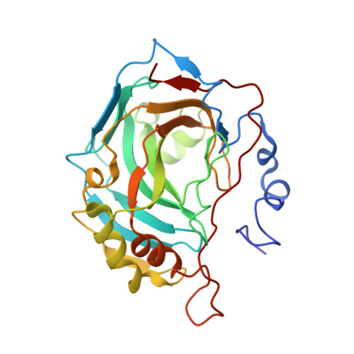
    JNJ-26990990 ((benzo[b]thien-3-yl)methyl)sulfamide, a sulfamide derivative structurally related to the antiepileptic drug zonisamide, was reported to be devoid of carbonic anhydrase (CA, EC 4.2.1.1) inhibitory properties. Here we report that JNJ-26990990 and its S,S-dioxide analog significantly inhibit six human (h) isoforms, hCA I, II, VII, IX, XII and XIV, involved in crucial physiological processes. Inhibition and X-ray crystallographic data for the binding of the two compounds to these enzymes show significant similarity with the zonisamide inhibitory pattern. These findings prompted us to reconsider the structural/pharmacological requirements for designing effective antiepileptics possessing zinc-binding groups of the sulfamide, sulfamate or sulfonamide type in their molecules.


  • Organizational Affiliation
    • Istituto di Biostrutture e Bioimmagini-CNR, via Mezzocannone 16, 80134 Napoli, Italy. gdesimon@unina.it claudiu.supuran@unifi.it.

Macromolecule Content 

  • Total Structure Weight: 30.06 kDa 
  • Atom Count: 2,352 
  • Modeled Residue Count: 258 
  • Deposited Residue Count: 260 
  • Unique protein chains: 1

Macromolecules

Find similar proteins by:|  3D Structure
Entity ID: 1
MoleculeChains  Sequence LengthOrganismDetailsImage
Carbonic anhydrase 2260Homo sapiensMutation(s): 0 
EC: 4.2.1.1 (PDB Primary Data), 4.2.1.69 (UniProt)
UniProt & NIH Common Fund Data Resources
Find proteins for P00918 (Homo sapiens)
Explore P00918 
Go to UniProtKB:  P00918
PHAROS:  P00918
GTEx:  ENSG00000104267 
Entity Groups
Sequence Clusters30% Identity50% Identity70% Identity90% Identity95% Identity100% Identity
UniProt GroupP00918
Sequence Annotations
Expand
Reference Sequence

Experimental Data & Validation

Experimental Data

  • Method: X-RAY DIFFRACTION
  • Resolution: 1.85 Å
  • R-Value Free:  0.193 (Depositor), 0.190 (DCC) 
  • R-Value Work:  0.162 (Depositor), 0.158 (DCC) 
Space Group: P 1 21 1
Unit Cell:
Length ( Å )Angle ( ˚ )
a = 42.006α = 90
b = 41.282β = 104.16
c = 71.981γ = 90
Software Package:
Software NamePurpose
d*TREKdata reduction
HKL-2000data scaling
CNSrefinement

Structure Validation

View Full Validation Report


Ligand Structure Quality Assessment 


Entry History 

Deposition Data

Revision History  (Full details and data files)

  • Version 1.0: 2016-05-18
    Type: Initial release
  • Version 1.1: 2016-06-01
    Changes: Database references
  • Version 1.2: 2024-01-10
    Changes: Data collection, Database references, Refinement description