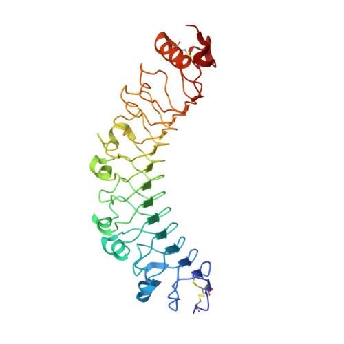
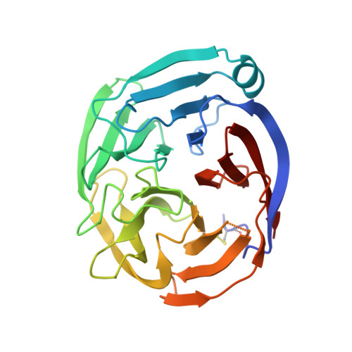
5CMN

FLRT3 LRR domain in complex with LPHN3 Olfactomedin domain


Experimental Data Snapshot

  • Method: X-RAY DIFFRACTION
  • Resolution: 3.60 Å
  • R-Value Free: 0.262 
  • R-Value Work: 0.195 
  • R-Value Observed: 0.199 

Starting Model: experimental
View more details

wwPDB Validation   3D Report Full Report


Ligand Structure Quality Assessment 


This is version 1.3 of the entry. See complete history


Literature

Structural Basis of Latrophilin-FLRT-UNC5 Interaction in Cell Adhesion.

Lu, Y.C.Nazarko, O.V.Sando, R.Salzman, G.S.Sudhof, T.C.Arac, D.

(2015) Structure 23: 1678-1691

  • DOI: https://doi.org/10.1016/j.str.2015.06.024
  • Primary Citation of Related Structures:  
    5CMN, 5CMP

  • PubMed Abstract: 

    Fibronectin leucine-rich repeat transmembrane proteins (FLRTs) are cell-adhesion molecules with emerging functions in cortical development and synapse formation. Their extracellular regions interact with latrophilins (LPHNs) to mediate synapse development, and with Uncoordinated-5 (UNC5)/netrin receptors to control the migration of neurons in the developing cortex. Here, we present the crystal structures of FLRT3 in isolation and in complex with LPHN3. The LPHN3/FLRT3 structure reveals that LPHN3 binds to FLRT3 at a site distinct from UNC5. Structure-based mutations specifically disrupt LPHN3/FLRT3 binding, but do not disturb their interactions with other proteins or their cell-membrane localization. Thus, they can be used as molecular tools to dissect the functions of FLRTs and LPHNs in vivo. Our results suggest that UNC5 and LPHN3 can simultaneously bind to FLRT3, forming a trimeric complex, and that FLRT3 may form transsynaptic complexes with both LPHN3 and UNC5. These findings provide molecular insights for understanding the role of cell-adhesion proteins in synapse function.


  • Organizational Affiliation

    Department of Biochemistry and Molecular Biology, The University of Chicago, 929 East 57(th) Street, GCIS W219, Chicago, IL 60637, USA.


Macromolecules
Find similar proteins by:  (by identity cutoff)  |  3D Structure
Entity ID: 1
MoleculeChains Sequence LengthOrganismDetailsImage
Leucine-rich repeat transmembrane protein FLRT3
A, B, C, D
333Homo sapiensMutation(s): 0 
Gene Names: FLRT3KIAA1469UNQ856/PRO1865
UniProt & NIH Common Fund Data Resources
Find proteins for Q9NZU0 (Homo sapiens)
Explore Q9NZU0 
Go to UniProtKB:  Q9NZU0
PHAROS:  Q9NZU0
GTEx:  ENSG00000125848 
Entity Groups  
Sequence Clusters30% Identity50% Identity70% Identity90% Identity95% Identity100% Identity
UniProt GroupQ9NZU0
Glycosylation
Glycosylation Sites: 1Go to GlyGen: Q9NZU0-1
Sequence Annotations
Expand
  • Reference Sequence
Find similar proteins by:  (by identity cutoff)  |  3D Structure
Entity ID: 2
MoleculeChains Sequence LengthOrganismDetailsImage
Latrophilin-3
E, F, G, H
261Homo sapiensMutation(s): 0 
Gene Names: LPHN3KIAA0768LEC3
UniProt & NIH Common Fund Data Resources
Find proteins for Q9HAR2 (Homo sapiens)
Explore Q9HAR2 
Go to UniProtKB:  Q9HAR2
PHAROS:  Q9HAR2
GTEx:  ENSG00000150471 
Entity Groups  
Sequence Clusters30% Identity50% Identity70% Identity90% Identity95% Identity100% Identity
UniProt GroupQ9HAR2
Sequence Annotations
Expand
  • Reference Sequence
Small Molecules
Ligands 2 Unique
IDChains Name / Formula / InChI Key2D Diagram3D Interactions
NAG
Query on NAG

Download Ideal Coordinates CCD File 
I [auth A],
J [auth B],
K [auth C],
L [auth D]
2-acetamido-2-deoxy-beta-D-glucopyranose
C8 H15 N O6
OVRNDRQMDRJTHS-FMDGEEDCSA-N
CA
Query on CA

Download Ideal Coordinates CCD File 
M [auth E],
N [auth F],
O [auth G],
P [auth H]
CALCIUM ION
Ca
BHPQYMZQTOCNFJ-UHFFFAOYSA-N
Experimental Data & Validation

Experimental Data

  • Method: X-RAY DIFFRACTION
  • Resolution: 3.60 Å
  • R-Value Free: 0.262 
  • R-Value Work: 0.195 
  • R-Value Observed: 0.199 
  • Space Group: P 43
Unit Cell:
Length ( Å )Angle ( ˚ )
a = 102.268α = 90
b = 102.268β = 90
c = 419.23γ = 90
Software Package:
Software NamePurpose
PHENIXrefinement
Cootmodel building
HKL-2000data scaling
HKL-2000data reduction
PHENIXphasing

Structure Validation

View Full Validation Report


Ligand Structure Quality Assessment 


Entry History 

Deposition Data

Revision History  (Full details and data files)

  • Version 1.0: 2015-08-12
    Type: Initial release
  • Version 1.1: 2015-09-16
    Changes: Database references
  • Version 1.2: 2020-07-29
    Type: Remediation
    Reason: Carbohydrate remediation
    Changes: Data collection, Database references, Derived calculations, Structure summary
  • Version 1.3: 2023-09-27
    Changes: Data collection, Database references, Refinement description, Structure summary