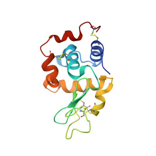
5C6J

Crystal Structure of Gadolinium derivative of HEWL solved using Free-Electron Laser radiation


Experimental Data Snapshot

  • Method: X-RAY DIFFRACTION
  • Resolution: 2.10 Å
  • R-Value Free: 0.272 
  • R-Value Work: 0.204 
  • R-Value Observed: 0.207 

wwPDB Validation   3D Report Full Report


This is version 1.3 of the entry. See complete history


Literature

Towards phasing using high X-ray intensity.

Galli, L.Son, S.K.Barends, T.R.White, T.A.Barty, A.Botha, S.Boutet, S.Caleman, C.Doak, R.B.Nanao, M.H.Nass, K.Shoeman, R.L.Timneanu, N.Santra, R.Schlichting, I.Chapman, H.N.

(2015) IUCrJ 2: 627-634

  • DOI: https://doi.org/10.1107/S2052252515014049
  • Primary Citation of Related Structures:  
    5C6I, 5C6J, 5C6L

  • PubMed Abstract: 

    X-ray free-electron lasers (XFELs) show great promise for macromolecular structure determination from sub-micrometre-sized crystals, using the emerging method of serial femtosecond crystallography. The extreme brightness of the XFEL radiation can multiply ionize most, if not all, atoms in a protein, causing their scattering factors to change during the pulse, with a preferential 'bleaching' of heavy atoms. This paper investigates the effects of electronic damage on experimental data collected from a Gd derivative of lysozyme microcrystals at different X-ray intensities, and the degree of ionization of Gd atoms is quantified from phased difference Fourier maps. A pattern sorting scheme is proposed to maximize the ionization contrast and the way in which the local electronic damage can be used for a new experimental phasing method is discussed.


  • Organizational Affiliation

    Center for Free-Electron Laser Science, Deutsches Elektronen-Synchrotron DESY , Notkestrasse 85, Hamburg, 22607, Germany ; Department of Physics, University of Hamburg , Luruper Chaussee 149, Hamburg, 22761, Germany.


Macromolecules
Find similar proteins by:  (by identity cutoff)  |  3D Structure
Entity ID: 1
MoleculeChains Sequence LengthOrganismDetailsImage
Lysozyme C129Gallus gallusMutation(s): 0 
EC: 3.2.1.17
UniProt
Find proteins for P00698 (Gallus gallus)
Explore P00698 
Go to UniProtKB:  P00698
Entity Groups  
Sequence Clusters30% Identity50% Identity70% Identity90% Identity95% Identity100% Identity
UniProt GroupP00698
Sequence Annotations
Expand
  • Reference Sequence
Small Molecules
Ligands 3 Unique
IDChains Name / Formula / InChI Key2D Diagram3D Interactions
DO3
Query on DO3

Download Ideal Coordinates CCD File 
B [auth A],
C [auth A]
10-((2R)-2-HYDROXYPROPYL)-1,4,7,10-TETRAAZACYCLODODECANE 1,4,7-TRIACETIC ACID
C17 H32 N4 O7
IQUHNCOJRJBMSU-CQSZACIVSA-N
GD
Query on GD

Download Ideal Coordinates CCD File 
H [auth A],
I [auth A]
GADOLINIUM ATOM
Gd
UIWYJDYFSGRHKR-UHFFFAOYSA-N
CL
Query on CL

Download Ideal Coordinates CCD File 
D [auth A],
E [auth A],
F [auth A],
G [auth A]
CHLORIDE ION
Cl
VEXZGXHMUGYJMC-UHFFFAOYSA-M
Experimental Data & Validation

Experimental Data

  • Method: X-RAY DIFFRACTION
  • Resolution: 2.10 Å
  • R-Value Free: 0.272 
  • R-Value Work: 0.204 
  • R-Value Observed: 0.207 
  • Space Group: P 43 21 2
Unit Cell:
Length ( Å )Angle ( ˚ )
a = 79.2α = 90
b = 79.2β = 90
c = 39.4γ = 90
Software Package:
Software NamePurpose
SCALAdata scaling
REFMACrefinement
PDB_EXTRACTdata extraction
ARPmodel building
SOLVEphasing
CrystFELdata reduction

Structure Validation

View Full Validation Report


Entry History 

Revision History  (Full details and data files)

  • Version 1.0: 2015-07-08
    Type: Initial release
  • Version 1.1: 2015-12-09
    Changes: Database references
  • Version 1.2: 2018-01-24
    Changes: Data collection
  • Version 1.3: 2018-11-14
    Changes: Data collection