5C1B | pdb_00005c1b

p97-delta709-728 in complex with a UFD1-SHP peptide


Experimental Data Snapshot

  • Method: X-RAY DIFFRACTION
  • Resolution: 3.08 Å
  • R-Value Free: 
    0.240 (Depositor), 0.251 (DCC) 
  • R-Value Work: 
    0.198 (Depositor), 0.210 (DCC) 
  • R-Value Observed: 
    0.200 (Depositor) 

Starting Model: experimental
View more details

wwPDB Validation 3D Report Full Report

Validation slider image for 5C1B

Ligand Structure Quality Assessment 


This is version 1.2 of the entry. See complete history

Literature

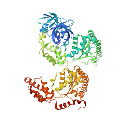
Characterization of an Additional Binding Surface on the p97 N-Terminal Domain Involved in Bipartite Cofactor Interactions.

Hanzelmann, P.Schindelin, H.

(2016) Structure 24: 140-147

  • DOI: https://doi.org/10.1016/j.str.2015.10.027
  • Primary Citation Related Structures: 
    5C1B

  • PubMed Abstract: 

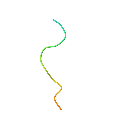
    The type II AAA ATPase p97 interacts with a large number of cofactors that regulate its function by recruiting it to different cellular pathways. Most of the cofactors interact with the N-terminal (N) domain of p97, either via ubiquitin-like domains or short linear binding motifs. While some linear binding motifs form α helices, another group features short stretches of unstructured hydrophobic sequences as found in the so-called SHP (BS1, binding segment 1) motif. Here we present the crystal structure of a SHP-binding motif in complex with p97, which reveals a so far uncharacterized binding site on the p97 N domain that is different from the conserved binding surface of all other known p97 cofactors. This finding explains how cofactors like UFD1/NPL4 and p47 can utilize a bipartite binding mechanism to interact simultaneously with the same p97 monomer via their ubiquitin-like domain and SHP motif.


  • Organizational Affiliation
    • Rudolf Virchow Center for Experimental Biomedicine, University of Würzburg, Josef-Schneider-Straße 2, 97080 Würzburg, Germany. Electronic address: petra.haenzelmann@virchow.uni-wuerzburg.de.

Macromolecule Content 

  • Total Structure Weight: 532.94 kDa 
  • Atom Count: 34,659 
  • Modeled Residue Count: 4,365 
  • Deposited Residue Count: 4,752 
  • Unique protein chains: 2

Macromolecules

Find similar proteins by:|  3D Structure
Entity ID: 1
MoleculeChains  Sequence LengthOrganismDetailsImage
Transitional endoplasmic reticulum ATPase
A, B, C, D, E
A, B, C, D, E, F
785Homo sapiensMutation(s): 0 
Gene Names: VCP
EC: 3.6.4.6
UniProt & NIH Common Fund Data Resources
Find proteins for P55072 (Homo sapiens)
Explore P55072 
Go to UniProtKB:  P55072
PHAROS:  P55072
GTEx:  ENSG00000165280 
Entity Groups
Sequence Clusters30% Identity50% Identity70% Identity90% Identity95% Identity100% Identity
UniProt GroupP55072
Sequence Annotations
Expand
Reference Sequence
Find similar proteins by:  Sequence   |   3D Structure  
Entity ID: 2
MoleculeChains  Sequence LengthOrganismDetailsImage
Ubiquitin fusion degradation protein 1 homologG [auth V],
H [auth U]
21Homo sapiensMutation(s): 0 
UniProt & NIH Common Fund Data Resources
Find proteins for Q92890 (Homo sapiens)
Explore Q92890 
Go to UniProtKB:  Q92890
PHAROS:  Q92890
GTEx:  ENSG00000070010 
Entity Groups
Sequence Clusters30% Identity50% Identity70% Identity90% Identity95% Identity100% Identity
UniProt GroupQ92890
Sequence Annotations
Expand
Reference Sequence

Small Molecules

Ligands 4 Unique
IDChains Name / Formula / InChI Key2D Diagram3D Interactions
AGS

Query on AGS


Download:Ideal Coordinates CCD File
CA [auth E]
DA [auth E]
HA [auth F]
I [auth A]
IA [auth F]
CA [auth E],
DA [auth E],
HA [auth F],
I [auth A],
IA [auth F],
J [auth A],
N [auth B],
O [auth B],
T [auth C],
U [auth C],
Y [auth D],
Z [auth D]
PHOSPHOTHIOPHOSPHORIC ACID-ADENYLATE ESTER
C10 H16 N5 O12 P3 S
NLTUCYMLOPLUHL-KQYNXXCUSA-N
GOL

Query on GOL


Download:Ideal Coordinates CCD File
MA [auth F],
NA [auth F]
GLYCEROL
C3 H8 O3
PEDCQBHIVMGVHV-UHFFFAOYSA-N
CL

Query on CL


Download:Ideal Coordinates CCD File
GA [auth E]
LA [auth F]
M [auth A]
R [auth B]
S [auth C]
GA [auth E],
LA [auth F],
M [auth A],
R [auth B],
S [auth C],
X [auth D]
CHLORIDE ION
Cl
VEXZGXHMUGYJMC-UHFFFAOYSA-M
MG

Query on MG


Download:Ideal Coordinates CCD File
AA [auth D]
BA [auth D]
EA [auth E]
FA [auth E]
JA [auth F]
AA [auth D],
BA [auth D],
EA [auth E],
FA [auth E],
JA [auth F],
K [auth A],
KA [auth F],
L [auth A],
P [auth B],
Q [auth B],
V [auth C],
W [auth C]
MAGNESIUM ION
Mg
JLVVSXFLKOJNIY-UHFFFAOYSA-N

Experimental Data & Validation

Experimental Data

  • Method: X-RAY DIFFRACTION
  • Resolution: 3.08 Å
  • R-Value Free:  0.240 (Depositor), 0.251 (DCC) 
  • R-Value Work:  0.198 (Depositor), 0.210 (DCC) 
  • R-Value Observed: 0.200 (Depositor) 
Space Group: P 21 21 21
Unit Cell:
Length ( Å )Angle ( ˚ )
a = 140.715α = 90
b = 180.664β = 90
c = 254.958γ = 90
Software Package:
Software NamePurpose
PHENIXrefinement
Aimlessdata scaling
MxCuBEdata collection
PDB_EXTRACTdata extraction
PHASERphasing
XDSdata reduction

Structure Validation

View Full Validation Report


Ligand Structure Quality Assessment 


Entry History 

Deposition Data

Revision History  (Full details and data files)

  • Version 1.0: 2016-01-13
    Type: Initial release
  • Version 1.1: 2016-01-20
    Changes: Database references
  • Version 1.2: 2024-01-10
    Changes: Data collection, Database references, Derived calculations, Refinement description