5B07

Lysozyme (denatured by DCl and refolded)

  • Classification: HYDROLASE
  • Organism(s): Gallus gallus
  • Mutation(s): No 

  • Deposited: 2015-10-28 Released: 2016-01-13 
  • Deposition Author(s): Kita, A., Morimoto, Y.
  • Funding Organization(s): Ministry of Education, Culture, Sports, Science and Technology (Japan)

Experimental Data Snapshot

  • Method: X-RAY DIFFRACTION
  • Resolution: 1.80 Å
  • R-Value Free: 0.187 
  • R-Value Work: 0.150 
  • R-Value Observed: 0.152 

wwPDB Validation   3D Report Full Report


This is version 1.2 of the entry. See complete history


Literature

An Effective Deuterium Exchange Method for Neutron Crystal Structure Analysis with Unfolding-Refolding Processes

Kita, A.Morimoto, Y.

(2016) Mol Biotechnol 58: 130-136

  • DOI: https://doi.org/10.1007/s12033-015-9908-8
  • Primary Citation of Related Structures:  
    5B05, 5B06, 5B07

  • PubMed Abstract: 

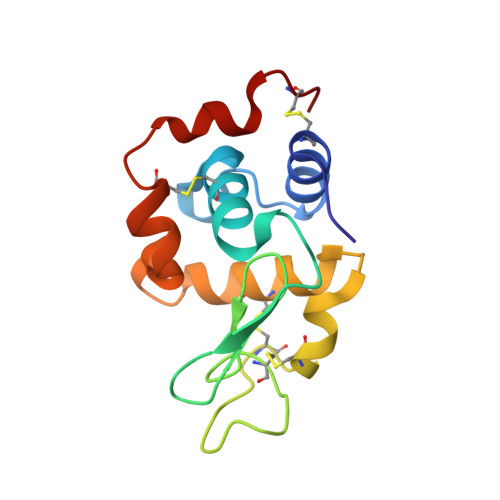
    A method of hydrogen/deuterium (H/D) exchange with an unfolding-refolding process has been applied to hen egg-white lysozyme (HWL), and accurate evaluation of its deuteration was carried out by time-of-flight mass spectroscopy. Neutron crystallography requires a suitable crystal with enough deuterium exchanged in the protein to decrease incoherent scattering from hydrogens. It is very expensive to prepare a fully deuterated protein, and therefore a simple H/D exchange technique is desirable for this purpose. Acid or base addition to protein solutions with heating effectively increased the number of deuterium up to more than 20 % of that of all hydrogen atoms, and refolded structures were determined by X-ray structure analysis at 1.8 Å resolution. Refolded HWL had increased deuterium content in its protein core and its native structure, determined at atomic resolution, was fully preserved.


  • Organizational Affiliation

    Division of the Quantum Beam Material Science, Research Reactor Institute, Kyoto University, Kumatori, Osaka, 590-0494, Japan. kita@rri.kyoto-u.ac.jp.


Macromolecules
Find similar proteins by:  (by identity cutoff)  |  3D Structure
Entity ID: 1
MoleculeChains Sequence LengthOrganismDetailsImage
Lysozyme C129Gallus gallusMutation(s): 0 
EC: 3.2.1.17
UniProt
Find proteins for P00698 (Gallus gallus)
Explore P00698 
Go to UniProtKB:  P00698
Entity Groups  
Sequence Clusters30% Identity50% Identity70% Identity90% Identity95% Identity100% Identity
UniProt GroupP00698
Sequence Annotations
Expand
  • Reference Sequence
Small Molecules
Ligands 3 Unique
IDChains Name / Formula / InChI Key2D Diagram3D Interactions
EDO
Query on EDO

Download Ideal Coordinates CCD File 
B [auth A],
C [auth A],
D [auth A],
E [auth A]
1,2-ETHANEDIOL
C2 H6 O2
LYCAIKOWRPUZTN-UHFFFAOYSA-N
CL
Query on CL

Download Ideal Coordinates CCD File 
N [auth A],
O [auth A],
P [auth A],
Q [auth A]
CHLORIDE ION
Cl
VEXZGXHMUGYJMC-UHFFFAOYSA-M
NA
Query on NA

Download Ideal Coordinates CCD File 
F [auth A]
G [auth A]
H [auth A]
I [auth A]
J [auth A]
F [auth A],
G [auth A],
H [auth A],
I [auth A],
J [auth A],
K [auth A],
L [auth A],
M [auth A]
SODIUM ION
Na
FKNQFGJONOIPTF-UHFFFAOYSA-N
Experimental Data & Validation

Experimental Data

  • Method: X-RAY DIFFRACTION
  • Resolution: 1.80 Å
  • R-Value Free: 0.187 
  • R-Value Work: 0.150 
  • R-Value Observed: 0.152 
  • Space Group: P 43 21 2
Unit Cell:
Length ( Å )Angle ( ˚ )
a = 78.816α = 90
b = 78.816β = 90
c = 36.8γ = 90
Software Package:
Software NamePurpose
REFMACrefinement
HKL-2000data reduction
HKL-2000data scaling
MOLREPphasing

Structure Validation

View Full Validation Report


Entry History & Funding Information

Deposition Data


Funding OrganizationLocationGrant Number
Ministry of Education, Culture, Sports, Science and Technology (Japan)Japan--

Revision History  (Full details and data files)

  • Version 1.0: 2016-01-13
    Type: Initial release
  • Version 1.1: 2016-02-10
    Changes: Database references
  • Version 1.2: 2023-11-08
    Changes: Author supporting evidence, Data collection, Database references, Derived calculations, Refinement description