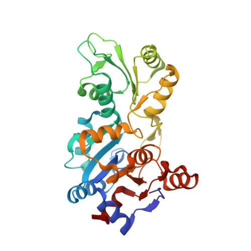
5AES

Crystal Structure of murine Chronophin (Pyridoxal Phosphate Phosphatase) in Complex with a PNP-derived Inhibitor


Experimental Data Snapshot

  • Method: X-RAY DIFFRACTION
  • Resolution: 2.75 Å
  • R-Value Free: 0.247 
  • R-Value Work: 0.190 
  • R-Value Observed: 0.192 

wwPDB Validation   3D Report Full Report


Ligand Structure Quality Assessment 


This is version 2.0 of the entry. See complete history


Literature

Synthesis of Hydrolysis-Resistant Pyridoxal 5'-Phosphate Analogs and Their Biochemical and X-Ray Crystallographic Characterization with the Pyridoxal Phosphatase Chronophin.

Knobloch, G.Jabari, N.Stadlbauer, S.Schindelin, H.Kohn, M.Gohla, A.

(2015) Bioorg Med Chem 23: 2819

  • DOI: https://doi.org/10.1016/j.bmc.2015.02.049
  • Primary Citation of Related Structures:  
    5AES

  • PubMed Abstract: 
  • A set of phosphonic acid derivatives (1-4) of pyridoxal 5'-phosphate (PLP) was synthesized and characterized biochemically using purified murine pyridoxal phosphatase (PDXP), also known as chronophin. The most promising compound 1 displayed primarily competitive PDXP inhibitory activity with an IC50 value of 79μM, which was in the range of the Km of the physiological substrate PLP ...

    A set of phosphonic acid derivatives (1-4) of pyridoxal 5'-phosphate (PLP) was synthesized and characterized biochemically using purified murine pyridoxal phosphatase (PDXP), also known as chronophin. The most promising compound 1 displayed primarily competitive PDXP inhibitory activity with an IC50 value of 79μM, which was in the range of the Km of the physiological substrate PLP. We also report the X-ray crystal structure of PDXP bound to compound 3, which we solved to 2.75Å resolution (PDB code 5AES). The co-crystal structure proves that compound 3 binds in the same orientation as PLP, and confirms the mode of inhibition to be competitive. Thus, we identify compound 1 as a PDXP phosphatase inhibitor. Our results suggest a strategy to design new, potent and selective PDXP inhibitors, which may be useful to increase the sensitivity of tumor cells to treatment with cytotoxic agents.


    Organizational Affiliation

    Institute of Pharmacology and Toxicology, University of Würzburg, Versbacher Strasse 9, D-97078 Würzburg, Germany; Rudolf Virchow Center for Experimental Biomedicine, University of Würzburg, Josef-Schneider-Strasse 2, D-97080 Würzburg, Germany. Electronic address: antje.gohla@virchow.uni-wuerzburg.de.


Macromolecules
Find similar proteins by:  (by identity cutoff)  |  3D Structure
Entity ID: 1
MoleculeChainsSequence LengthOrganismDetailsImage
PYRIDOXAL PHOSPHATE PHOSPHATASEA, B293Mus musculusMutation(s): 0 
Gene Names: PdxpCinPlpPlpp
EC: 3.1.3.74 (PDB Primary Data), 3.1.3.16 (UniProt)
UniProt & NIH Common Fund Data Resources
Find proteins for P60487 (Mus musculus)
Explore P60487 
Go to UniProtKB:  P60487
IMPC:  MGI:1919282
Entity Groups  
Sequence Clusters30% Identity50% Identity70% Identity90% Identity95% Identity100% Identity
UniProt GroupP60487
Protein Feature View
Expand
  • Reference Sequence
Small Molecules
Ligands 3 Unique
IDChainsName / Formula / InChI Key2D Diagram3D Interactions
5B0
Query on 5B0

Download Ideal Coordinates CCD File 
D [auth A],
F [auth B]
{2-[5-hydroxy-4-(hydroxymethyl)-6-methylpyridin-3-yl]ethyl}phosphonic acid
C9 H14 N O5 P
JWJVWJVYBWLXDN-UHFFFAOYSA-N
 Ligand Interaction
GOL
Query on GOL

Download Ideal Coordinates CCD File 
G [auth B]GLYCEROL
C3 H8 O3
PEDCQBHIVMGVHV-UHFFFAOYSA-N
 Ligand Interaction
MG
Query on MG

Download Ideal Coordinates CCD File 
C [auth A],
E [auth B]
MAGNESIUM ION
Mg
JLVVSXFLKOJNIY-UHFFFAOYSA-N
 Ligand Interaction
Binding Affinity Annotations 
IDSourceBinding Affinity
5B0 BindingDB:  5AES IC50: 1.30e+6 (nM) from 1 assay(s)
Experimental Data & Validation

Experimental Data

  • Method: X-RAY DIFFRACTION
  • Resolution: 2.75 Å
  • R-Value Free: 0.247 
  • R-Value Work: 0.190 
  • R-Value Observed: 0.192 
  • Space Group: I 2 3
Unit Cell:
Length ( Å )Angle ( ˚ )
a = 166.81α = 90
b = 166.81β = 90
c = 166.81γ = 90
Software Package:
Software NamePurpose
PHENIXrefinement
iMOSFLMdata reduction
SCALAdata scaling
PHASERphasing

Structure Validation

View Full Validation Report


Ligand Structure Quality Assessment 


Entry History 

Deposition Data

Revision History  (Full details and data files)

  • Version 1.0: 2015-04-01
    Type: Initial release
  • Version 1.1: 2015-04-15
    Changes: Atomic model, Other
  • Version 1.2: 2015-06-10
    Changes: Database references
  • Version 2.0: 2017-06-28
    Changes: Atomic model, Data collection