4PQG

Crystal structure of the pneumococcal O-GlcNAc transferase GtfA in complex with UDP and GlcNAc


Experimental Data Snapshot

  • Method: X-RAY DIFFRACTION
  • Resolution: 2.00 Å
  • R-Value Free: 0.248 
  • R-Value Work: 0.199 
  • R-Value Observed: 0.201 

wwPDB Validation   3D Report Full Report


Ligand Structure Quality Assessment 


This is version 1.2 of the entry. See complete history


Literature

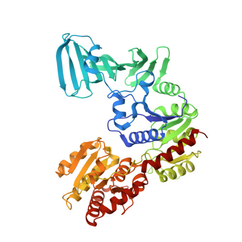
Structure of a Novel O-Linked N-Acetyl-d-glucosamine (O-GlcNAc) Transferase, GtfA, Reveals Insights into the Glycosylation of Pneumococcal Serine-rich Repeat Adhesins.

Shi, W.W.Jiang, Y.L.Zhu, F.Yang, Y.H.Shao, Q.Y.Yang, H.B.Ren, Y.M.Wu, H.Chen, Y.Zhou, C.Z.

(2014) J Biol Chem 289: 20898-20907

  • DOI: https://doi.org/10.1074/jbc.M114.581934
  • Primary Citation of Related Structures:  
    4PQG

  • PubMed Abstract: 
  • Protein glycosylation catalyzed by the O-GlcNAc transferase (OGT) plays a critical role in various biological processes. In Streptococcus pneumoniae, the core enzyme GtfA and co-activator GtfB form an OGT complex to glycosylate the serine-rich repeat (SRR) of adhesin PsrP (pneumococcal serine-rich repeat protein), which is involved in the infection and pathogenesis ...

    Protein glycosylation catalyzed by the O-GlcNAc transferase (OGT) plays a critical role in various biological processes. In Streptococcus pneumoniae, the core enzyme GtfA and co-activator GtfB form an OGT complex to glycosylate the serine-rich repeat (SRR) of adhesin PsrP (pneumococcal serine-rich repeat protein), which is involved in the infection and pathogenesis. Here we report the 2.0 Å crystal structure of GtfA, revealing a β-meander add-on domain beyond the catalytic domain. It represents a novel add-on domain, which is distinct from the all-α-tetratricopeptide repeats in the only two structure-known OGTs. Structural analyses combined with binding assays indicate that this add-on domain contributes to forming an active GtfA-GtfB complex and recognizing the acceptor protein. In addition, the in vitro glycosylation system enables us to map the O-linkages to the serine residues within the first SRR of PsrP. These findings suggest that fusion with an add-on domain might be a universal mechanism for diverse OGTs that recognize varying acceptor proteins/peptides.


    Organizational Affiliation

    University of Ottawa, Ottawa Institute of Systems Biology, 451 Smyth Rd., Roger Guindon Hall, Ottawa, ON K1H 8M5, Canada.


Macromolecules
Find similar proteins by:  (by identity cutoff)  |  3D Structure
Entity ID: 1
MoleculeChainsSequence LengthOrganismDetailsImage
Glycosyltransferase Gtf1
A, B
511Streptococcus pneumoniae TIGR4Mutation(s): 0 
Gene Names: gtf1SP_1758gtfA
EC: 2.4.1
UniProt
Find proteins for A0A0H2URG7 (Streptococcus pneumoniae serotype 4 (strain ATCC BAA-334 / TIGR4))
Explore A0A0H2URG7 
Go to UniProtKB:  A0A0H2URG7
Entity Groups  
Sequence Clusters30% Identity50% Identity70% Identity90% Identity95% Identity100% Identity
UniProt GroupA0A0H2URG7
Protein Feature View
Expand
  • Reference Sequence
Small Molecules
Ligands 2 Unique
IDChainsName / Formula / InChI Key2D Diagram3D Interactions
UDP
Query on UDP

Download Ideal Coordinates CCD File 
C [auth A],
E [auth B]
URIDINE-5'-DIPHOSPHATE
C9 H14 N2 O12 P2
XCCTYIAWTASOJW-XVFCMESISA-N
 Ligand Interaction
NAG
Query on NAG

Download Ideal Coordinates CCD File 
D [auth A],
F [auth B]
2-acetamido-2-deoxy-beta-D-glucopyranose
C8 H15 N O6
OVRNDRQMDRJTHS-FMDGEEDCSA-N
 Ligand Interaction
Experimental Data & Validation

Experimental Data

  • Method: X-RAY DIFFRACTION
  • Resolution: 2.00 Å
  • R-Value Free: 0.248 
  • R-Value Work: 0.199 
  • R-Value Observed: 0.201 
  • Space Group: P 21 21 21
Unit Cell:
Length ( Å )Angle ( ˚ )
a = 71.24α = 90
b = 125.63β = 90
c = 127.04γ = 90
Software Package:
Software NamePurpose
ADSCdata collection
SHELXSphasing
REFMACrefinement
MOSFLMdata reduction
SCALAdata scaling

Structure Validation

View Full Validation Report


Ligand Structure Quality Assessment 


Entry History 

Deposition Data

Revision History  (Full details and data files)

  • Version 1.0: 2014-06-18
    Type: Initial release
  • Version 1.1: 2014-08-06
    Changes: Database references
  • Version 1.2: 2020-07-29
    Type: Remediation
    Reason: Carbohydrate remediation
    Changes: Data collection, Database references, Derived calculations, Structure summary