4HAA

Structure of Ribonuclease Binase Glu43Ala/Phe81Ala Mutant


Experimental Data Snapshot

  • Method: X-RAY DIFFRACTION
  • Resolution: 1.90 Å
  • R-Value Free: 0.205 
  • R-Value Work: 0.177 
  • R-Value Observed: 0.179 

wwPDB Validation   3D Report Full Report


This is version 1.3 of the entry. See complete history


Literature

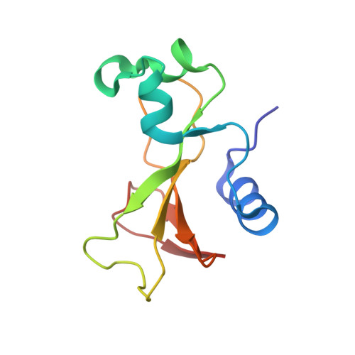
Structure and functional studies of the ribonuclease binase Glu43Ala/Phe81Ala mutant.

Mitkevich, V.A.Schulga, A.A.Trofimov, A.A.Dorovatovskii, P.V.Goncharuk, D.A.Tkach, E.N.Makarov, A.A.Polyakov, K.M.

(2013) Acta Crystallogr D Biol Crystallogr 69: 991-996

  • DOI: https://doi.org/10.1107/S0907444913004046
  • Primary Citation of Related Structures:  
    4HAA

  • PubMed Abstract: 

    Ribonuclease from Bacillus intermedius (binase) is a small basic protein with antitumour activity. The three-dimensional structure of the binase mutant form Glu43Ala/Phe81Ala was determined at 1.98 Å resolution and its functional properties, such as the kinetic parameters characterizing the hydrolysis of polyinosinic acid and cytotoxicity towards Kasumi-1 cells, were investigated. In all crystal structures of binase studied previously the characteristic dimer is present, with the active site of one subunit being blocked owing to interactions within the dimer. In contrast to this, the new mutant form is not dimeric in the crystal. The catalytic efficiency of the mutant form is increased 1.7-fold and its cytotoxic properties are enhanced compared with the wild-type enzyme.


  • Organizational Affiliation

    Engelhardt Institute of Molecular Biology, Russian Academy of Sciences, ul. Vavilova 32, Moscow 119991, Russian Federation.


Macromolecules
Find similar proteins by:  (by identity cutoff)  |  3D Structure
Entity ID: 1
MoleculeChains Sequence LengthOrganismDetailsImage
Ribonuclease
A, B, C, D
109Bacillus intermediusMutation(s): 2 
EC: 3.1.27
UniProt
Find proteins for P00649 (Bacillus intermedius)
Explore P00649 
Go to UniProtKB:  P00649
Entity Groups  
Sequence Clusters30% Identity50% Identity70% Identity90% Identity95% Identity100% Identity
UniProt GroupP00649
Sequence Annotations
Expand
  • Reference Sequence
Experimental Data & Validation

Experimental Data

  • Method: X-RAY DIFFRACTION
  • Resolution: 1.90 Å
  • R-Value Free: 0.205 
  • R-Value Work: 0.177 
  • R-Value Observed: 0.179 
  • Space Group: P 43
Unit Cell:
Length ( Å )Angle ( ˚ )
a = 50.36α = 90
b = 50.36β = 90
c = 196.12γ = 90
Software Package:
Software NamePurpose
REFMACrefinement
XDSdata reduction
XSCALEdata scaling
MOLREPphasing

Structure Validation

View Full Validation Report


Entry History 

Deposition Data

Revision History  (Full details and data files)

  • Version 1.0: 2012-10-17
    Type: Initial release
  • Version 1.1: 2013-05-29
    Changes: Database references
  • Version 1.2: 2014-02-05
    Changes: Database references
  • Version 1.3: 2023-09-20
    Changes: Data collection, Database references, Refinement description