4GFH

Topoisomerase II-DNA-AMPPNP complex


Experimental Data Snapshot

  • Method: X-RAY DIFFRACTION
  • Resolution: 4.41 Å
  • R-Value Free: 0.275 
  • R-Value Work: 0.239 
  • R-Value Observed: 0.241 

wwPDB Validation   3D Report Full Report


Ligand Structure Quality Assessment 


This is version 1.2 of the entry. See complete history


Literature

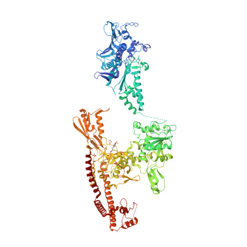
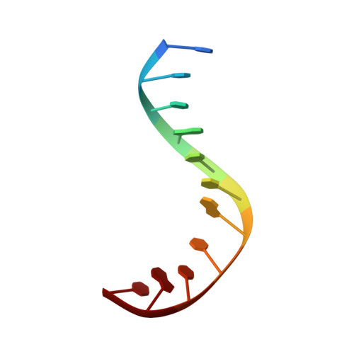
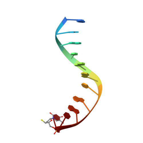
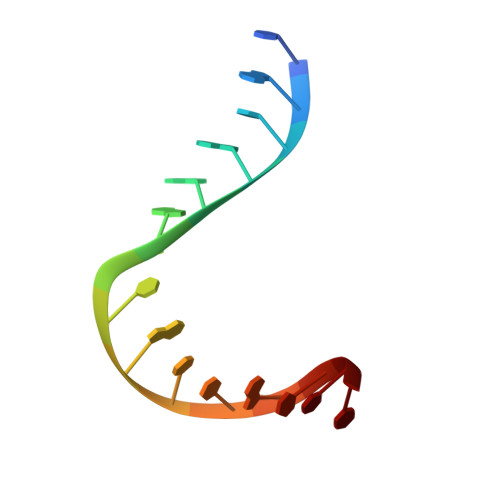
Structure of a topoisomerase II-DNA-nucleotide complex reveals a new control mechanism for ATPase activity.

Schmidt, B.H.Osheroff, N.Berger, J.M.

(2012) Nat Struct Mol Biol 19: 1147-1154

  • DOI: https://doi.org/10.1038/nsmb.2388
  • Primary Citation of Related Structures:  
    4GFH

  • PubMed Abstract: 
  • Type IIA topoisomerases control DNA supercoiling and disentangle chromosomes through a complex ATP-dependent strand-passage mechanism. Although a general framework exists for type IIA topoisomerase function, the architecture of the full-length enzyme has remained undefined ...

    Type IIA topoisomerases control DNA supercoiling and disentangle chromosomes through a complex ATP-dependent strand-passage mechanism. Although a general framework exists for type IIA topoisomerase function, the architecture of the full-length enzyme has remained undefined. Here we present the structure of a fully catalytic Saccharomyces cerevisiae topoisomerase II homodimer complexed with DNA and a nonhydrolyzable ATP analog. The enzyme adopts a domain-swapped configuration wherein the ATPase domain of one protomer sits atop the nucleolytic region of its partner subunit. This organization produces an unexpected interaction between bound DNA and a conformational transducing element in the ATPase domain, which we show is critical for both DNA-stimulated ATP hydrolysis and global topoisomerase activity. Our data indicate that the ATPase domains pivot about each other to ensure unidirectional strand passage and that this state senses bound DNA to promote ATP turnover and enzyme reset.


    Organizational Affiliation

    Department of Molecular and Cell Biology, University of California, Berkeley, Berkeley, California, USA.


Macromolecules

Find similar proteins by:  (by identity cutoff)  |  3D Structure
Entity ID: 1
MoleculeChainsSequence LengthOrganismDetailsImage
DNA topoisomerase 2
A, F
1,177Saccharomyces cerevisiae S288CMutation(s): 0 
Gene Names: TOP2TOR3YNL088WN2244
EC: 5.99.1.3 (PDB Primary Data), 5.6.2.2 (UniProt)
UniProt
Find proteins for P06786 (Saccharomyces cerevisiae (strain ATCC 204508 / S288c))
Explore P06786 
Go to UniProtKB:  P06786
Entity Groups  
Sequence Clusters30% Identity50% Identity70% Identity90% Identity95% Identity100% Identity
UniProt GroupP06786
Protein Feature View
Expand
  • Reference Sequence

Find similar nucleic acids by:  Sequence   |   3D Structure  

Entity ID: 2
MoleculeChainsLengthOrganismImage
DNA (5'-D(P*CP*CP*TP*AP*CP*TP*GP*CP*TP*AP*C)-3')
B, G
11N/A
Protein Feature View
Expand
  • Reference Sequence

Find similar nucleic acids by:  Sequence   |   3D Structure  

Entity ID: 3
MoleculeChainsLengthOrganismImage
DNA (5'-D(*CP*GP*CP*GP*GP*TP*AP*GP*CP*AP*GP*TP*AP*GP*G)-3')
C, H
15N/A
Protein Feature View
Expand
  • Reference Sequence

Find similar nucleic acids by:  Sequence   |   3D Structure  

Entity ID: 4
MoleculeChainsLengthOrganismImage
DNA (5'-D(P*GP*GP*AP*TP*GP*AP*CP*GP*AP*TP*(TSP))-3')
D, I
11N/A
Protein Feature View
Expand
  • Reference Sequence

Find similar nucleic acids by:  Sequence   |   3D Structure  

Entity ID: 5
MoleculeChainsLengthOrganismImage
DNA (5'-D(*CP*GP*CP*GP*AP*AP*TP*CP*GP*TP*CP*AP*TP*CP*C)-3')
E, J
15N/A
Protein Feature View
Expand
  • Reference Sequence
Small Molecules
Ligands 2 Unique
IDChainsName / Formula / InChI Key2D Diagram3D Interactions
ANP
Query on ANP

Download Ideal Coordinates CCD File 
L [auth A],
N [auth F]
PHOSPHOAMINOPHOSPHONIC ACID-ADENYLATE ESTER
C10 H17 N6 O12 P3
PVKSNHVPLWYQGJ-KQYNXXCUSA-N
 Ligand Interaction
MG
Query on MG

Download Ideal Coordinates CCD File 
K [auth A],
M [auth F]
MAGNESIUM ION
Mg
JLVVSXFLKOJNIY-UHFFFAOYSA-N
 Ligand Interaction
Modified Residues  1 Unique
IDChainsTypeFormula2D DiagramParent
PTR
Query on PTR
A, F
L-PEPTIDE LINKINGC9 H12 N O6 PTYR
Experimental Data & Validation

Experimental Data

  • Method: X-RAY DIFFRACTION
  • Resolution: 4.41 Å
  • R-Value Free: 0.275 
  • R-Value Work: 0.239 
  • R-Value Observed: 0.241 
  • Space Group: P 21 21 21
Unit Cell:
Length ( Å )Angle ( ˚ )
a = 169.135α = 90
b = 169.884β = 90
c = 169.212γ = 90
Software Package:
Software NamePurpose
ADSCdata collection
PHASERphasing
PHENIXrefinement
HKL-2000data reduction
HKL-2000data scaling

Structure Validation

View Full Validation Report


Ligand Structure Quality Assessment 


Entry History 

Deposition Data

Revision History  (Full details and data files)

  • Version 1.0: 2012-10-03
    Type: Initial release
  • Version 1.1: 2012-12-12
    Changes: Database references
  • Version 1.2: 2019-07-31
    Changes: Data collection, Derived calculations