4ZRL | pdb_00004zrl

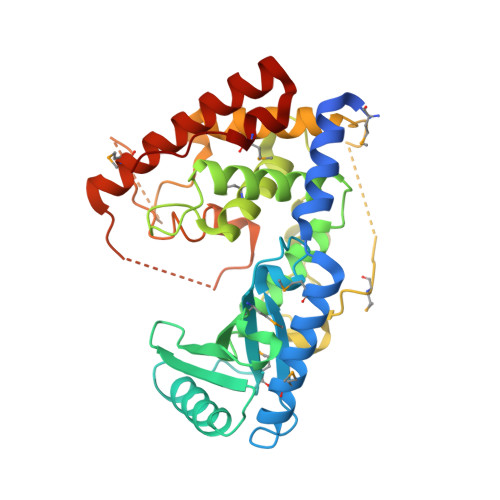
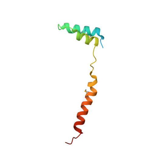
Structure of the non canonical Poly(A) polymerase complex GLD-2 - GLD-3


Experimental Data Snapshot

  • Method: X-RAY DIFFRACTION
  • Resolution: 2.28 Å
  • R-Value Free: 
    0.231 (Depositor), 0.230 (DCC) 
  • R-Value Work: 
    0.187 (Depositor), 0.190 (DCC) 
  • R-Value Observed: 
    0.189 (Depositor) 

wwPDB Validation   3D Report Full Report


This is version 2.1 of the entry. See complete history


Literature

Structural basis for the activation of the C. elegans noncanonical cytoplasmic poly(A)-polymerase GLD-2 by GLD-3.

Nakel, K.Bonneau, F.Eckmann, C.R.Conti, E.

(2015) Proc Natl Acad Sci U S A 112: 8614-8619

  • DOI: https://doi.org/10.1073/pnas.1504648112
  • Primary Citation of Related Structures:  
    4ZRL

  • PubMed Abstract: 

    The Caenorhabditis elegans germ-line development defective (GLD)-2-GLD-3 complex up-regulates the expression of genes required for meiotic progression. GLD-2-GLD-3 acts by extending the short poly(A) tail of germ-line-specific mRNAs, switching them from a dormant state into a translationally active state. GLD-2 is a cytoplasmic noncanonical poly(A) polymerase that lacks the RNA-binding domain typical of the canonical nuclear poly(A)-polymerase Pap1. The activity of C. elegans GLD-2 in vivo and in vitro depends on its association with the multi-K homology (KH) domain-containing protein, GLD-3, a homolog of Bicaudal-C. We have identified a minimal polyadenylation complex that includes the conserved nucleotidyl-transferase core of GLD-2 and the N-terminal domain of GLD-3, and determined its structure at 2.3-Å resolution. The structure shows that the N-terminal domain of GLD-3 does not fold into the predicted KH domain but wraps around the catalytic domain of GLD-2. The picture that emerges from the structural and biochemical data are that GLD-3 activates GLD-2 both indirectly by stabilizing the enzyme and directly by contributing positively charged residues near the RNA-binding cleft. The RNA-binding cleft of GLD-2 has distinct structural features compared with the poly(A)-polymerases Pap1 and Trf4. Consistently, GLD-2 has distinct biochemical properties: It displays unusual specificity in vitro for single-stranded RNAs with at least one adenosine at the 3' end. GLD-2 thus appears to have evolved specialized nucleotidyl-transferase properties that match the 3' end features of dormant cytoplasmic mRNAs.


  • Organizational Affiliation

    Department of Structural Cell Biology, Max Planck Institute of Biochemistry, Am Klopferspitz 18, D-82152 Martinsried, Germany;


Macromolecules
Find similar proteins by:  (by identity cutoff)  |  3D Structure
Entity ID: 1
MoleculeChains Sequence LengthOrganismDetailsImage
Poly(A) RNA polymerase gld-2364Caenorhabditis elegansMutation(s): 0 
Gene Names: gld-2ZC308.1
EC: 2.7.7.19
UniProt
Find proteins for O17087 (Caenorhabditis elegans)
Explore O17087 
Go to UniProtKB:  O17087
Entity Groups  
Sequence Clusters30% Identity50% Identity70% Identity90% Identity95% Identity100% Identity
UniProt GroupO17087
Sequence Annotations
Expand
  • Reference Sequence
Find similar proteins by:  (by identity cutoff)  |  3D Structure
Entity ID: 2
MoleculeChains Sequence LengthOrganismDetailsImage
Defective in germ line development protein 377Caenorhabditis elegansMutation(s): 0 
Gene Names: gld-3T07F8.3
UniProt
Find proteins for Q95ZK7 (Caenorhabditis elegans)
Explore Q95ZK7 
Go to UniProtKB:  Q95ZK7
Entity Groups  
Sequence Clusters30% Identity50% Identity70% Identity90% Identity95% Identity100% Identity
UniProt GroupQ95ZK7
Sequence Annotations
Expand
  • Reference Sequence
Small Molecules
Ligands 1 Unique
IDChains Name / Formula / InChI Key2D Diagram3D Interactions
CL
Query on CL

Download Ideal Coordinates CCD File 
C [auth A]CHLORIDE ION
Cl
VEXZGXHMUGYJMC-UHFFFAOYSA-M
Modified Residues  1 Unique
IDChains TypeFormula2D DiagramParent
MSE
Query on MSE
A
L-PEPTIDE LINKINGC5 H11 N O2 SeMET
Experimental Data & Validation

Experimental Data

  • Method: X-RAY DIFFRACTION
  • Resolution: 2.28 Å
  • R-Value Free:  0.231 (Depositor), 0.230 (DCC) 
  • R-Value Work:  0.187 (Depositor), 0.190 (DCC) 
  • R-Value Observed: 0.189 (Depositor) 
Space Group: P 21 21 21
Unit Cell:
Length ( Å )Angle ( ˚ )
a = 50.655α = 90
b = 89.367β = 90
c = 94.666γ = 90
Software Package:
Software NamePurpose
PHENIXrefinement
XDSdata reduction
SHELXDEphasing
Cootmodel building
XDSdata scaling

Structure Validation

View Full Validation Report


Entry History & Funding Information

Deposition Data


Funding OrganizationLocationGrant Number
German Research FoundationGermanyBU2451/1-2
EC | European Research Council (ERC): Elena Conti ERC Advanced Investigator Grant294371
German Research FoundationSFB646, SFB1035, GRK1721, FOR1680
German Research FoundationFOR855, EC369/3-1

Revision History  (Full details and data files)

  • Version 1.0: 2015-07-08
    Type: Initial release
  • Version 1.1: 2015-07-15
    Changes: Database references
  • Version 1.2: 2015-07-22
    Changes: Database references
  • Version 1.3: 2016-07-13
    Changes: Data collection
  • Version 2.0: 2017-09-06
    Changes: Advisory, Atomic model, Author supporting evidence, Refinement description
  • Version 2.1: 2024-11-20
    Changes: Data collection, Database references, Structure summary