4Z9O

Crystal Structure of human GGT1


Experimental Data Snapshot

  • Method: X-RAY DIFFRACTION
  • Resolution: 2.30 Å
  • R-Value Free: 0.201 
  • R-Value Work: 0.152 
  • R-Value Observed: 0.154 

wwPDB Validation   3D Report Full Report


Ligand Structure Quality Assessment 


This is version 1.5 of the entry. See complete history


Literature

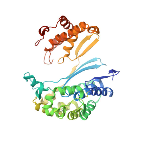
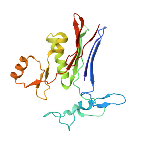
Human gamma-Glutamyl Transpeptidase 1: STRUCTURES OF THE FREE ENZYME, INHIBITOR-BOUND TETRAHEDRAL TRANSITION STATES, AND GLUTAMATE-BOUND ENZYME REVEAL NOVEL MOVEMENT WITHIN THE ACTIVE SITE DURING CATALYSIS.

Terzyan, S.S.Burgett, A.W.Heroux, A.Smith, C.A.Mooers, B.H.Hanigan, M.H.

(2015) J Biol Chem 290: 17576-17586

  • DOI: https://doi.org/10.1074/jbc.M115.659680
  • Primary Citation of Related Structures:  
    4Z9O, 4ZBK, 4ZC6, 4ZCG

  • PubMed Abstract: 

    γ-Glutamyl transpeptidase 1 (GGT1) is a cell surface, N-terminal nucleophile hydrolase that cleaves glutathione and other γ-glutamyl compounds. GGT1 expression is essential in cysteine homeostasis, and its induction has been implicated in the pathology of asthma, reperfusion injury, and cancer. In this study, we report four new crystal structures of human GGT1 (hGGT1) that show conformational changes within the active site as the enzyme progresses from the free enzyme to inhibitor-bound tetrahedral transition states and finally to the glutamate-bound structure prior to the release of this final product of the reaction. The structure of the apoenzyme shows flexibility within the active site. The serine-borate-bound hGGT1 crystal structure demonstrates that serine-borate occupies the active site of the enzyme, resulting in an enzyme-inhibitor complex that replicates the enzyme's tetrahedral intermediate/transition state. The structure of GGsTop-bound hGGT1 reveals its interactions with the enzyme and why neutral phosphonate diesters are more potent inhibitors than monoanionic phosphonates. These structures are the first structures for any eukaryotic GGT that include a molecule in the active site covalently bound to the catalytic Thr-381. The glutamate-bound structure shows the conformation of the enzyme prior to release of the final product and reveals novel information regarding the displacement of the main chain atoms that form the oxyanion hole and movement of the lid loop region when the active site is occupied. These data provide new insights into the mechanism of hGGT1-catalyzed reactions and will be invaluable in the development of new classes of hGGT1 inhibitors for therapeutic use.


  • Organizational Affiliation

    From the Macromolecular Crystallography Laboratory, Department of Biochemistry and Molecular Biology, University of Oklahoma Health Sciences Center, Oklahoma City, Oklahoma 73104.


Macromolecules
Find similar proteins by:  (by identity cutoff)  |  3D Structure
Entity ID: 1
MoleculeChains Sequence LengthOrganismDetailsImage
Gamma-glutamyltranspeptidase 1353Homo sapiensMutation(s): 1 
Gene Names: GGT1GGT
EC: 2.3.2.2 (PDB Primary Data), 3.4.19.13 (PDB Primary Data), 3.4.19.14 (PDB Primary Data)
UniProt & NIH Common Fund Data Resources
Find proteins for P19440 (Homo sapiens)
Explore P19440 
Go to UniProtKB:  P19440
PHAROS:  P19440
GTEx:  ENSG00000100031 
Entity Groups  
Sequence Clusters30% Identity50% Identity70% Identity90% Identity95% Identity100% Identity
UniProt GroupP19440
Sequence Annotations
Expand
  • Reference Sequence
Find similar proteins by:  (by identity cutoff)  |  3D Structure
Entity ID: 2
MoleculeChains Sequence LengthOrganismDetailsImage
Gamma-glutamyltranspeptidase 1189Homo sapiensMutation(s): 0 
Gene Names: GGT1GGT
EC: 2.3.2.2 (PDB Primary Data), 3.4.19.13 (PDB Primary Data), 3.4.19.14 (PDB Primary Data)
UniProt & NIH Common Fund Data Resources
Find proteins for P19440 (Homo sapiens)
Explore P19440 
Go to UniProtKB:  P19440
PHAROS:  P19440
GTEx:  ENSG00000100031 
Entity Groups  
Sequence Clusters30% Identity50% Identity70% Identity90% Identity95% Identity100% Identity
UniProt GroupP19440
Sequence Annotations
Expand
  • Reference Sequence
Small Molecules
Ligands 4 Unique
IDChains Name / Formula / InChI Key2D Diagram3D Interactions
NAG
Query on NAG

Download Ideal Coordinates CCD File 
C [auth A]
D [auth A]
E [auth A]
F [auth A]
G [auth A]
C [auth A],
D [auth A],
E [auth A],
F [auth A],
G [auth A],
J [auth B]
2-acetamido-2-deoxy-beta-D-glucopyranose
C8 H15 N O6
OVRNDRQMDRJTHS-FMDGEEDCSA-N
MES
Query on MES

Download Ideal Coordinates CCD File 
H [auth A],
K [auth B]
2-(N-MORPHOLINO)-ETHANESULFONIC ACID
C6 H13 N O4 S
SXGZJKUKBWWHRA-UHFFFAOYSA-N
CL
Query on CL

Download Ideal Coordinates CCD File 
I [auth A],
L [auth B]
CHLORIDE ION
Cl
VEXZGXHMUGYJMC-UHFFFAOYSA-M
NA
Query on NA

Download Ideal Coordinates CCD File 
M [auth B]SODIUM ION
Na
FKNQFGJONOIPTF-UHFFFAOYSA-N
Experimental Data & Validation

Experimental Data

  • Method: X-RAY DIFFRACTION
  • Resolution: 2.30 Å
  • R-Value Free: 0.201 
  • R-Value Work: 0.152 
  • R-Value Observed: 0.154 
  • Space Group: C 2 2 21
Unit Cell:
Length ( Å )Angle ( ˚ )
a = 105.583α = 90
b = 125.288β = 90
c = 103.988γ = 90
Software Package:
Software NamePurpose
REFMACrefinement
HKL-2000data reduction
HKL-2000data scaling
PDB_EXTRACTdata extraction
REFMACphasing

Structure Validation

View Full Validation Report


Ligand Structure Quality Assessment 


Entry History & Funding Information

Deposition Data


Funding OrganizationLocationGrant Number
National Institutes of Health/National Institute of General Medical Sciences (NIH/NIGMS)United StatesP20GM103640

Revision History  (Full details and data files)

  • Version 1.0: 2015-06-03
    Type: Initial release
  • Version 1.1: 2015-06-10
    Changes: Database references
  • Version 1.2: 2015-07-22
    Changes: Database references
  • Version 1.3: 2017-09-27
    Changes: Author supporting evidence, Database references, Derived calculations, Source and taxonomy
  • Version 1.4: 2019-12-25
    Changes: Author supporting evidence
  • Version 1.5: 2020-07-29
    Type: Remediation
    Reason: Carbohydrate remediation
    Changes: Data collection, Derived calculations, Structure summary