4X33

Structure of the Elongator cofactor complex Kti11/Kti13 at 1.45A


Experimental Data Snapshot

  • Method: X-RAY DIFFRACTION
  • Resolution: 1.45 Å
  • R-Value Free: 0.176 
  • R-Value Work: 0.150 
  • R-Value Observed: 0.151 

wwPDB Validation   3D Report Full Report


This is version 1.2 of the entry. See complete history


Literature

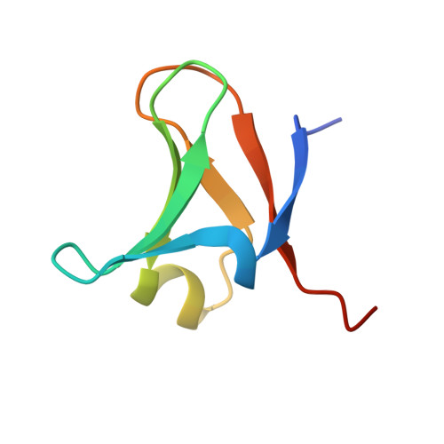
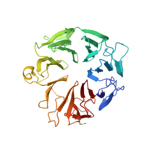
Structure of the Elongator cofactor complex Kti11/Kti13 provides insight into the role of Kti13 in Elongator-dependent tRNA modification.

Kolaj-Robin, O.McEwen, A.G.Cavarelli, J.Seraphin, B.

(2015) FEBS J 282: 819-833

  • DOI: https://doi.org/10.1111/febs.13199

  • PubMed Abstract: 

    Modification of wobble uridines of many eukaryotic tRNAs requires the Elongator complex, a highly conserved six-subunit eukaryotic protein assembly, as well as the Killer toxin-insensitive (Kti) proteins 11-14. Kti11 was additionally shown to be implicated in the biosynthesis of diphthamide, a post-translationally modified histidine of translation elongation factor 2. Recent data indicate that iron-bearing Kti11 functions as an electron donor to the [4Fe-4S] cluster of radical S-Adenosylmethionine enzymes, triggering the subsequent radical reaction. We show here that recombinant yeast Kti11 forms a stable 1 : 1 complex with Kti13. To obtain insights into the function of this heterodimer, the Kti11/Kti13 complex was purified to homogeneity, crystallized, and its structure determined at 1.45 Å resolution. The importance of several residues mediating complex formation was confirmed by mutagenesis. Kti13 adopts a fold characteristic of RCC1-like proteins. The seven-bladed β-propeller consists of a unique mixture of four- and three-stranded blades. In the complex, Kti13 orients Kti11 and restricts access to its electron-carrying iron atom, constraining the electron transfer capacity of Kti11. Based on these findings, we propose a role for Kti13, and discuss the possible functional implications of complex formation.


  • Organizational Affiliation

    Equipe Labellisée La Ligue, Institut de Génétique et de Biologie Moléculaire et Cellulaire, Centre National de Recherche Scientifique UMR 7104/Institut National de Santé et de Recherche Médicale U964/Université de Strasbourg, Illkirch, France.


Macromolecules
Find similar proteins by:  (by identity cutoff)  |  3D Structure
Entity ID: 1
MoleculeChains Sequence LengthOrganismDetailsImage
Diphthamide biosynthesis protein 363Saccharomyces cerevisiae S288CMutation(s): 0 
Gene Names: KTI11DPH3YBL071W-A
UniProt
Find proteins for Q3E840 (Saccharomyces cerevisiae (strain ATCC 204508 / S288c))
Explore Q3E840 
Go to UniProtKB:  Q3E840
Entity Groups  
Sequence Clusters30% Identity50% Identity70% Identity90% Identity95% Identity100% Identity
UniProt GroupQ3E840
Sequence Annotations
Expand
  • Reference Sequence
Find similar proteins by:  (by identity cutoff)  |  3D Structure
Entity ID: 2
MoleculeChains Sequence LengthOrganismDetailsImage
Protein ATS1333Saccharomyces cerevisiae S288CMutation(s): 0 
Gene Names: ATS1YAL020CFUN28YAL006
UniProt
Find proteins for P31386 (Saccharomyces cerevisiae (strain ATCC 204508 / S288c))
Explore P31386 
Go to UniProtKB:  P31386
Entity Groups  
Sequence Clusters30% Identity50% Identity70% Identity90% Identity95% Identity100% Identity
UniProt GroupP31386
Sequence Annotations
Expand
  • Reference Sequence
Small Molecules
Ligands 4 Unique
IDChains Name / Formula / InChI Key2D Diagram3D Interactions
DXE
Query on DXE

Download Ideal Coordinates CCD File 
L [auth B]1,2-DIMETHOXYETHANE
C4 H10 O2
XTHFKEDIFFGKHM-UHFFFAOYSA-N
FE
Query on FE

Download Ideal Coordinates CCD File 
C [auth A]FE (III) ION
Fe
VTLYFUHAOXGGBS-UHFFFAOYSA-N
CL
Query on CL

Download Ideal Coordinates CCD File 
D [auth A]
E [auth A]
F [auth A]
G [auth B]
H [auth B]
D [auth A],
E [auth A],
F [auth A],
G [auth B],
H [auth B],
I [auth B],
J [auth B],
K [auth B]
CHLORIDE ION
Cl
VEXZGXHMUGYJMC-UHFFFAOYSA-M
MG
Query on MG

Download Ideal Coordinates CCD File 
M [auth B]MAGNESIUM ION
Mg
JLVVSXFLKOJNIY-UHFFFAOYSA-N
Modified Residues  1 Unique
IDChains TypeFormula2D DiagramParent
CSO
Query on CSO
B
L-PEPTIDE LINKINGC3 H7 N O3 SCYS
Experimental Data & Validation

Experimental Data

  • Method: X-RAY DIFFRACTION
  • Resolution: 1.45 Å
  • R-Value Free: 0.176 
  • R-Value Work: 0.150 
  • R-Value Observed: 0.151 
  • Space Group: C 2 2 21
Unit Cell:
Length ( Å )Angle ( ˚ )
a = 49.79α = 90
b = 89.009β = 90
c = 163.53γ = 90
Software Package:
Software NamePurpose
Aimlessdata scaling
BUSTER-TNTrefinement
PDB_EXTRACTdata extraction
XDSdata reduction

Structure Validation

View Full Validation Report


Entry History & Funding Information

Deposition Data


Funding OrganizationLocationGrant Number
France--

Revision History  (Full details and data files)

  • Version 1.0: 2015-01-21
    Type: Initial release
  • Version 1.1: 2015-02-04
    Changes: Database references
  • Version 1.2: 2015-03-11
    Changes: Database references