4WSS | pdb_00004wss

The crystal structure of hemagglutinin form A/chicken/New York/14677-13/1998 in complex with LSTa


Experimental Data Snapshot

  • Method: X-RAY DIFFRACTION
  • Resolution: 2.80 Å
  • R-Value Free: 
    0.254 (Depositor), 0.251 (DCC) 
  • R-Value Work: 
    0.220 (Depositor), 0.221 (DCC) 
  • R-Value Observed: 
    0.221 (Depositor) 

wwPDB Validation 3D Report Full Report

Validation slider image for 4WSS

Ligand Structure Quality Assessment 


This is version 2.2 of the entry. See complete history

Literature

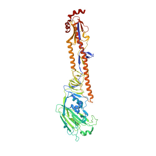
Structure and receptor binding preferences of recombinant hemagglutinins from avian and human h6 and h10 influenza a virus subtypes.

Yang, H.Carney, P.J.Chang, J.C.Villanueva, J.M.Stevens, J.

(2015) J Virol 89: 4612-4623

  • DOI: https://doi.org/10.1128/JVI.03456-14
  • Primary Citation Related Structures: 
    4WSR, 4WSS, 4WST, 4WSU, 4WSV, 4WSW, 4WSX

  • PubMed Abstract: 

    During 2013, three new avian influenza A virus subtypes, A(H7N9), A(H6N1), and A(H10N8), resulted in human infections. While the A(H7N9) virus resulted in a significant epidemic in China across 19 provinces and municipalities, both A(H6N1) and A(H10N8) viruses resulted in only a few human infections. This study focuses on the major surface glycoprotein hemagglutinins from both of these novel human viruses. The detailed structural and glycan microarray analyses presented here highlight the idea that both A(H6N1) and A(H10N8) virus hemagglutinins retain a strong avian receptor binding preference and thus currently pose a low risk for sustained human infections. Human infections with zoonotic influenza virus subtypes continue to be a great public health concern. We report detailed structural analysis and glycan microarray data for recombinant hemagglutinins from A(H6N1) and A(H10N8) viruses, isolated from human infections in 2013, and compare them with hemagglutinins of avian origin. This is the first structural report of an H6 hemagglutinin, and our results should further the understanding of these viruses and provide useful information to aid in the continuous surveillance of these zoonotic influenza viruses.


  • Organizational Affiliation
    • Influenza Division, National Center for Immunization and Respiratory Diseases, Centers for Disease Control and Prevention, Atlanta, Georgia, USA.

Macromolecule Content 

  • Total Structure Weight: 351.66 kDa 
  • Atom Count: 23,442 
  • Modeled Residue Count: 2,914 
  • Deposited Residue Count: 3,084 
  • Unique protein chains: 1

Macromolecules

Find similar proteins by:|  3D Structure
Entity ID: 1
MoleculeChains  Sequence LengthOrganismDetailsImage
Hemagglutinin514Influenza A virus (A/chicken/New York/14677-13/1998(H6N2))Mutation(s): 0 
Gene Names: HA
UniProt
Find proteins for Q0A3A5 (Influenza A virus)
Explore Q0A3A5 
Go to UniProtKB:  Q0A3A5
Entity Groups
Sequence Clusters30% Identity50% Identity70% Identity90% Identity95% Identity100% Identity
UniProt GroupQ0A3A5
Glycosylation
Glycosylation Sites: 1
Sequence Annotations
Expand
Reference Sequence

Oligosaccharides

Help  
Entity ID: 2
MoleculeChains Length2D Diagram GlycosylationD Interactions
2-acetamido-2-deoxy-beta-D-glucopyranose-(1-4)-2-acetamido-2-deoxy-beta-D-glucopyranose
G, J
2N-Glycosylation
Glycosylation Resources
GlyTouCan: G42666HT
GlyCosmos: G42666HT
GlyGen: G42666HT
Entity ID: 3
MoleculeChains Length2D Diagram GlycosylationD Interactions
N-acetyl-alpha-neuraminic acid-(2-3)-beta-D-galactopyranose-(1-4)-2-acetamido-2-deoxy-beta-D-glucopyranose
H, I, K
3N/A
Glycosylation Resources
GlyTouCan: G00065MO
GlyCosmos: G00065MO
GlyGen: G00065MO

Experimental Data & Validation

Experimental Data

  • Method: X-RAY DIFFRACTION
  • Resolution: 2.80 Å
  • R-Value Free:  0.254 (Depositor), 0.251 (DCC) 
  • R-Value Work:  0.220 (Depositor), 0.221 (DCC) 
  • R-Value Observed: 0.221 (Depositor) 
Space Group: C 1 2 1
Unit Cell:
Length ( Å )Angle ( ˚ )
a = 254.967α = 90
b = 134.779β = 112.2
c = 122.622γ = 90
Software Package:
Software NamePurpose
REFMACrefinement
HKL-2000data reduction
SCALEPACKdata scaling
PHASERphasing

Structure Validation

View Full Validation Report


Ligand Structure Quality Assessment 


Entry History 

Deposition Data

Revision History  (Full details and data files)

  • Version 1.0: 2015-02-25
    Type: Initial release
  • Version 1.1: 2015-04-01
    Changes: Database references
  • Version 2.0: 2020-07-29
    Type: Remediation
    Reason: Carbohydrate remediation
    Changes: Advisory, Atomic model, Data collection, Database references, Derived calculations, Source and taxonomy, Structure summary
  • Version 2.1: 2023-12-27
    Changes: Data collection, Database references, Derived calculations, Refinement description, Structure summary
  • Version 2.2: 2024-10-09
    Changes: Structure summary