4WG7

Room-temperature crystal structure of lysozyme determined by serial synchrotron crystallography using a nano focused beam.


Experimental Data Snapshot

  • Method: X-RAY DIFFRACTION
  • Resolution: 1.70 Å
  • R-Value Free: 0.239 
  • R-Value Work: 0.204 
  • R-Value Observed: 0.205 

wwPDB Validation   3D Report Full Report


This is version 1.4 of the entry. See complete history


Literature

Raster-scanning serial protein crystallography using micro- and nano-focused synchrotron beams.

Coquelle, N.Brewster, A.S.Kapp, U.Shilova, A.Weinhausen, B.Burghammer, M.Colletier, J.P.

(2015) Acta Crystallogr D Biol Crystallogr 71: 1184-1196

  • DOI: https://doi.org/10.1107/S1399004715004514
  • Primary Citation of Related Structures:  
    4WG1, 4WG7, 4WL6, 4WL7

  • PubMed Abstract: 

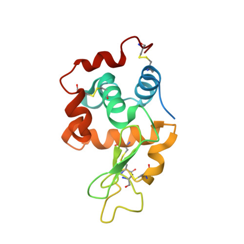
    High-resolution structural information was obtained from lysozyme microcrystals (20 µm in the largest dimension) using raster-scanning serial protein crystallography on micro- and nano-focused beamlines at the ESRF. Data were collected at room temperature (RT) from crystals sandwiched between two silicon nitride wafers, thereby preventing their drying, while limiting background scattering and sample consumption. In order to identify crystal hits, new multi-processing and GUI-driven Python-based pre-analysis software was developed, named NanoPeakCell, that was able to read data from a variety of crystallographic image formats. Further data processing was carried out using CrystFEL, and the resultant structures were refined to 1.7 Å resolution. The data demonstrate the feasibility of RT raster-scanning serial micro- and nano-protein crystallography at synchrotrons and validate it as an alternative approach for the collection of high-resolution structural data from micro-sized crystals. Advantages of the proposed approach are its thriftiness, its handling-free nature, the reduced amount of sample required, the adjustable hit rate, the high indexing rate and the minimization of background scattering.


  • Organizational Affiliation

    Université Grenoble Alpes, IBS, 38044 Grenoble, France.


Macromolecules
Find similar proteins by:  (by identity cutoff)  |  3D Structure
Entity ID: 1
MoleculeChains Sequence LengthOrganismDetailsImage
Lysozyme C147Gallus gallusMutation(s): 0 
EC: 3.2.1.17
UniProt
Find proteins for P00698 (Gallus gallus)
Explore P00698 
Go to UniProtKB:  P00698
Entity Groups  
Sequence Clusters30% Identity50% Identity70% Identity90% Identity95% Identity100% Identity
UniProt GroupP00698
Sequence Annotations
Expand
  • Reference Sequence
Small Molecules
Ligands 1 Unique
IDChains Name / Formula / InChI Key2D Diagram3D Interactions
CL
Query on CL

Download Ideal Coordinates CCD File 
B [auth A]CHLORIDE ION
Cl
VEXZGXHMUGYJMC-UHFFFAOYSA-M
Experimental Data & Validation

Experimental Data

  • Method: X-RAY DIFFRACTION
  • Resolution: 1.70 Å
  • R-Value Free: 0.239 
  • R-Value Work: 0.204 
  • R-Value Observed: 0.205 
  • Space Group: P 43 21 2
Unit Cell:
Length ( Å )Angle ( ˚ )
a = 77.99α = 90
b = 77.99β = 90
c = 38.51γ = 90
Software Package:
Software NamePurpose
PHENIXrefinement
PHASERphasing
CrystFELdata scaling
CrystFELdata reduction

Structure Validation

View Full Validation Report


Entry History & Funding Information

Deposition Data


Funding OrganizationLocationGrant Number
Fondation France AlzheimerFrance--

Revision History  (Full details and data files)

  • Version 1.0: 2015-05-06
    Type: Initial release
  • Version 1.1: 2015-05-13
    Changes: Database references
  • Version 1.2: 2015-05-20
    Changes: Database references
  • Version 1.3: 2016-07-27
    Changes: Database references
  • Version 1.4: 2024-01-10
    Changes: Data collection, Database references, Refinement description