4W8F | pdb_00004w8f

Crystal structure of the dynein motor domain in the AMPPNP-bound state


Experimental Data Snapshot

  • Method: X-RAY DIFFRACTION
  • Resolution: 3.54 Å
  • R-Value Free: 
    0.262 (Depositor), 0.225 (DCC) 
  • R-Value Work: 
    0.228 (Depositor), 0.232 (DCC) 
  • R-Value Observed: 
    0.230 (Depositor) 

wwPDB Validation 3D Report Full Report

Validation slider image for 4W8F

Ligand Structure Quality Assessment 


This is version 1.4 of the entry. See complete history

Literature

Allosteric communication in the Dynein motor domain.

Bhabha, G.Cheng, H.C.Zhang, N.Moeller, A.Liao, M.Speir, J.A.Cheng, Y.Vale, R.D.

(2014) Cell 159: 857-868

  • DOI: https://doi.org/10.1016/j.cell.2014.10.018
  • Primary Citation Related Structures: 
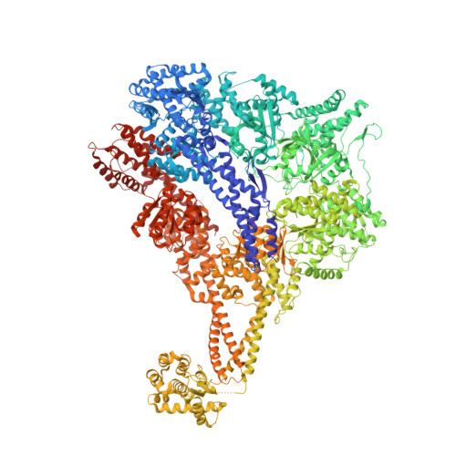
    4W8F

  • PubMed Abstract: 

    Dyneins power microtubule motility using ring-shaped, AAA-containing motor domains. Here, we report X-ray and electron microscopy (EM) structures of yeast dynein bound to different ATP analogs, which collectively provide insight into the roles of dynein's two major ATPase sites, AAA1 and AAA3, in the conformational change mechanism. ATP binding to AAA1 triggers a cascade of conformational changes that propagate to all six AAA domains and cause a large movement of the "linker," dynein's mechanical element. In contrast to the role of AAA1 in driving motility, nucleotide transitions in AAA3 gate the transmission of conformational changes between AAA1 and the linker, suggesting that AAA3 acts as a regulatory switch. Further structural and mutational studies also uncover a role for the linker in regulating the catalytic cycle of AAA1. Together, these results reveal how dynein's two major ATP-binding sites initiate and modulate conformational changes in the motor domain during motility.


  • Organizational Affiliation
    • Howard Hughes Medical Institute and the Department of Cellular and Molecular Pharmacology, University of California, San Francisco, San Francisco, CA 94158, USA.

Macromolecule Content 

  • Total Structure Weight: 613.88 kDa 
  • Atom Count: 42,393 
  • Modeled Residue Count: 5,217 
  • Deposited Residue Count: 5,322 
  • Unique protein chains: 1

Macromolecules

Find similar proteins by:|  3D Structure
Entity ID: 1
MoleculeChains  Sequence LengthOrganismDetailsImage
Dynein heavy chain lysozyme chimera
A, B
2,661Saccharomyces cerevisiae S288CTequatrovirus T4Mutation(s): 1 
Gene Names: DYN1DHC1YKR054C
EC: 3.2.1.17
UniProt
Find proteins for P00720 (Enterobacteria phage T4)
Explore P00720 
Go to UniProtKB:  P00720
Find proteins for P36022 (Saccharomyces cerevisiae (strain ATCC 204508 / S288c))
Explore P36022 
Go to UniProtKB:  P36022
Entity Groups
Sequence Clusters30% Identity50% Identity70% Identity90% Identity95% Identity100% Identity
UniProt GroupsP00720P36022
Sequence Annotations
Expand
Reference Sequence

Small Molecules

Ligands 2 Unique
IDChains Name / Formula / InChI Key2D Diagram3D Interactions
ANP

Query on ANP


Download:Ideal Coordinates CCD File
C [auth A]
D [auth A]
E [auth A]
F [auth A]
H [auth B]
C [auth A],
D [auth A],
E [auth A],
F [auth A],
H [auth B],
I [auth B],
J [auth B],
K [auth B]
PHOSPHOAMINOPHOSPHONIC ACID-ADENYLATE ESTER
C10 H17 N6 O12 P3
PVKSNHVPLWYQGJ-KQYNXXCUSA-N
MG

Query on MG


Download:Ideal Coordinates CCD File
G [auth A],
L [auth B]
MAGNESIUM ION
Mg
JLVVSXFLKOJNIY-UHFFFAOYSA-N

Experimental Data & Validation

Experimental Data

  • Method: X-RAY DIFFRACTION
  • Resolution: 3.54 Å
  • R-Value Free:  0.262 (Depositor), 0.225 (DCC) 
  • R-Value Work:  0.228 (Depositor), 0.232 (DCC) 
  • R-Value Observed: 0.230 (Depositor) 
Space Group: P 1 21 1
Unit Cell:
Length ( Å )Angle ( ˚ )
a = 137.73α = 90
b = 154.38β = 96.59
c = 177.55γ = 90
Software Package:
Software NamePurpose
PHENIXrefinement

Structure Validation

View Full Validation Report


Ligand Structure Quality Assessment 


Entry History 

& Funding Information

Deposition Data


Funding OrganizationLocationGrant Number
National Institutes of Health/National Institute of General Medical Sciences (NIH/NIGMS)United StatesR01GM09731

Revision History  (Full details and data files)

  • Version 1.0: 2014-11-12
    Type: Initial release
  • Version 1.1: 2014-12-10
    Changes: Database references
  • Version 1.2: 2017-09-20
    Changes: Author supporting evidence, Derived calculations, Other, Source and taxonomy, Structure summary
  • Version 1.3: 2019-12-25
    Changes: Author supporting evidence
  • Version 1.4: 2023-12-27
    Changes: Data collection, Database references, Derived calculations, Refinement description