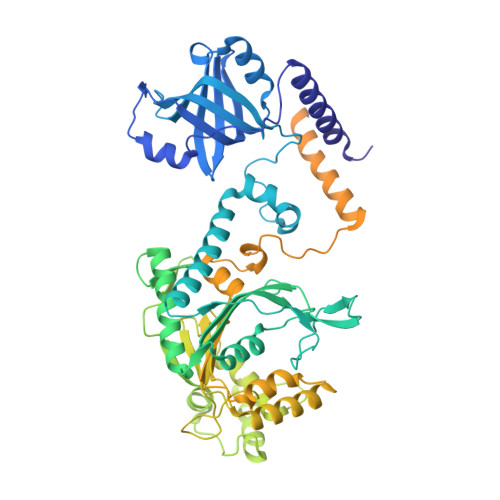
4UP9

Crystal structure of Entamoeba histolytica lysyl-tRNA synthetase in complex with ATP


Experimental Data Snapshot

  • Method: X-RAY DIFFRACTION
  • Resolution: 3.05 Å
  • R-Value Free: 0.263 
  • R-Value Work: 0.216 
  • R-Value Observed: 0.219 

wwPDB Validation   3D Report Full Report


Ligand Structure Quality Assessment 


This is version 1.4 of the entry. See complete history


Literature

Crystal Structures of Entamoeba Histolytica Lysyl-tRNA Synthetase Reveal Conformational Changes Upon Lysine Binding and a Specific Helix Bundle Domain.

Bonnefond, L.Castro De Moura, M.Ribas De Pouplana, L.Nureki, O.

(2014) FEBS Lett 588: 4478

  • DOI: https://doi.org/10.1016/j.febslet.2014.10.019
  • Primary Citation of Related Structures:  
    4UP7, 4UP8, 4UP9, 4UPA

  • PubMed Abstract: 

    The class II lysyl-tRNA synthetases (KRS) are conserved aminoacyl-tRNA synthetases that attach lysine to the cognate tRNA in a two-step mechanism. The enzyme from the parasitic protozoan Entamoeba histolytica was crystallized in the presence of small ligands to generate snapshots of the lysine-adenylate formation. The residues involved in lysine activation are highly conserved and the active site closes around the lysyl-adenylate, as observed in bacterial KRS. The Entamoeba EMAPII-like polypeptide is not resolved in the crystals, but another Entamoeba-specific insertion could be modeled as a small helix bundle that may contribute to tRNA binding through interaction with the tRNA hinge.


  • Organizational Affiliation

    Department of Biophysics and Biochemistry, Graduate School of Science, The University of Tokyo, 2-11-16, Yayoi, Bunkyo, Tokyo 113-0032, Japan. lbonnefond@unistra.fr


Macromolecules
Find similar proteins by:  (by identity cutoff)  |  3D Structure
Entity ID: 1
MoleculeChains Sequence LengthOrganismDetailsImage
LYSINE--TRNA LIGASE777Entamoeba histolyticaMutation(s): 1 
EC: 6.1.1.6
UniProt
Find proteins for C4M7X2 (Entamoeba histolytica (strain ATCC 30459 / HM-1:IMSS / ABRM))
Explore C4M7X2 
Go to UniProtKB:  C4M7X2
Entity Groups  
Sequence Clusters30% Identity50% Identity70% Identity90% Identity95% Identity100% Identity
UniProt GroupC4M7X2
Sequence Annotations
Expand
  • Reference Sequence
Small Molecules
Ligands 1 Unique
IDChains Name / Formula / InChI Key2D Diagram3D Interactions
ATP
Query on ATP

Download Ideal Coordinates CCD File 
B [auth A]ADENOSINE-5'-TRIPHOSPHATE
C10 H16 N5 O13 P3
ZKHQWZAMYRWXGA-KQYNXXCUSA-N
Experimental Data & Validation

Experimental Data

  • Method: X-RAY DIFFRACTION
  • Resolution: 3.05 Å
  • R-Value Free: 0.263 
  • R-Value Work: 0.216 
  • R-Value Observed: 0.219 
  • Space Group: P 62
Unit Cell:
Length ( Å )Angle ( ˚ )
a = 155.551α = 90
b = 155.551β = 90
c = 92.926γ = 120
Software Package:
Software NamePurpose
PHENIXrefinement
XDSdata reduction
Aimlessdata scaling
PHASERphasing

Structure Validation

View Full Validation Report


Ligand Structure Quality Assessment 


Entry History 

Deposition Data

Revision History  (Full details and data files)

  • Version 1.0: 2014-10-29
    Type: Initial release
  • Version 1.1: 2014-11-05
    Changes: Database references
  • Version 1.2: 2014-11-26
    Changes: Database references
  • Version 1.3: 2014-12-10
    Changes: Database references
  • Version 1.4: 2024-01-10
    Changes: Data collection, Database references, Derived calculations, Other, Refinement description