4U3J

TOG2:alpha/beta-tubulin complex


Experimental Data Snapshot

  • Method: X-RAY DIFFRACTION
  • Resolution: 2.81 Å
  • R-Value Free: 0.259 
  • R-Value Work: 0.218 
  • R-Value Observed: 0.220 

wwPDB Validation   3D Report Full Report


Ligand Structure Quality Assessment 


This is version 1.3 of the entry. See complete history


Literature

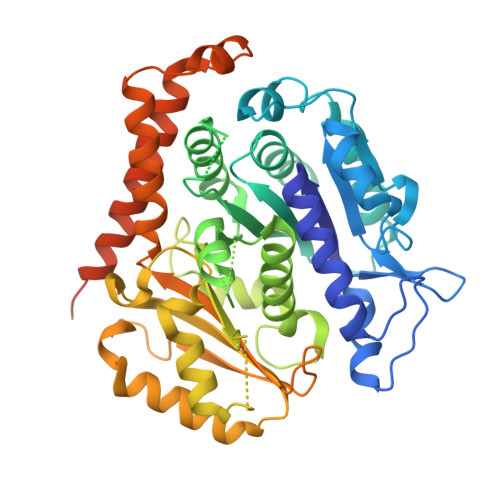
A tethered delivery mechanism explains the catalytic action of a microtubule polymerase.

Ayaz, P.Munyoki, S.Geyer, E.A.Piedra, F.A.Vu, E.S.Bromberg, R.Otwinowski, Z.Grishin, N.V.Brautigam, C.A.Rice, L.M.

(2014) Elife 3: e03069-e03069

  • DOI: https://doi.org/10.7554/eLife.03069
  • Primary Citation of Related Structures:  
    4U3J

  • PubMed Abstract: 
  • Stu2p/XMAP215 proteins are essential microtubule polymerases that use multiple αβ-tubulin-interacting TOG domains to bind microtubule plus ends and catalyze fast microtubule growth. We report here the structure of the TOG2 domain from Stu2p bound to yeast αβ-tubulin ...

    Stu2p/XMAP215 proteins are essential microtubule polymerases that use multiple αβ-tubulin-interacting TOG domains to bind microtubule plus ends and catalyze fast microtubule growth. We report here the structure of the TOG2 domain from Stu2p bound to yeast αβ-tubulin. Like TOG1, TOG2 binds selectively to a fully 'curved' conformation of αβ-tubulin, incompatible with a microtubule lattice. We also show that TOG1-TOG2 binds non-cooperatively to two αβ-tubulins. Preferential interactions between TOGs and fully curved αβ-tubulin that cannot exist elsewhere in the microtubule explain how these polymerases localize to the extreme microtubule end. We propose that these polymerases promote elongation because their linked TOG domains concentrate unpolymerized αβ-tubulin near curved subunits already bound at the microtubule end. This tethering model can explain catalyst-like behavior and also predicts that the polymerase action changes the configuration of the microtubule end.


    Organizational Affiliation

    Department of Biophysics, UT Southwestern Medical Center, Dallas, United States Department of Biochemistry, UT Southwestern Medical Center, Dallas, United States Luke.Rice@UTSouthwestern.edu.


Macromolecules
Find similar proteins by:  (by identity cutoff)  |  3D Structure
Entity ID: 1
MoleculeChainsSequence LengthOrganismDetailsImage
Tubulin alpha-1 chainA447Saccharomyces cerevisiaeMutation(s): 0 
Gene Names: TUB1
UniProt
Find proteins for P09733 (Saccharomyces cerevisiae (strain ATCC 204508 / S288c))
Explore P09733 
Go to UniProtKB:  P09733
Entity Groups  
Sequence Clusters30% Identity50% Identity70% Identity90% Identity95% Identity100% Identity
UniProt GroupP09733
Protein Feature View
Expand
  • Reference Sequence
Find similar proteins by:  (by identity cutoff)  |  3D Structure
Entity ID: 2
MoleculeChainsSequence LengthOrganismDetailsImage
Tubulin beta chainB463Saccharomyces cerevisiaeMutation(s): 2 
Gene Names: TUB2
UniProt
Find proteins for P02557 (Saccharomyces cerevisiae (strain ATCC 204508 / S288c))
Explore P02557 
Go to UniProtKB:  P02557
Entity Groups  
Sequence Clusters30% Identity50% Identity70% Identity90% Identity95% Identity100% Identity
UniProt GroupP02557
Protein Feature View
Expand
  • Reference Sequence
Find similar proteins by:  (by identity cutoff)  |  3D Structure
Entity ID: 3
MoleculeChainsSequence LengthOrganismDetailsImage
Protein STU2C249Saccharomyces cerevisiaeMutation(s): 0 
Gene Names: STU2
UniProt
Find proteins for P46675 (Saccharomyces cerevisiae (strain ATCC 204508 / S288c))
Explore P46675 
Go to UniProtKB:  P46675
Entity Groups  
Sequence Clusters30% Identity50% Identity70% Identity90% Identity95% Identity100% Identity
UniProt GroupP46675
Protein Feature View
Expand
  • Reference Sequence
Small Molecules
Ligands 2 Unique
IDChainsName / Formula / InChI Key2D Diagram3D Interactions
GTP
Query on GTP

Download Ideal Coordinates CCD File 
E [auth A],
G [auth B]
GUANOSINE-5'-TRIPHOSPHATE
C10 H16 N5 O14 P3
XKMLYUALXHKNFT-UUOKFMHZSA-N
 Ligand Interaction
MG
Query on MG

Download Ideal Coordinates CCD File 
D [auth A],
F [auth B]
MAGNESIUM ION
Mg
JLVVSXFLKOJNIY-UHFFFAOYSA-N
 Ligand Interaction
Experimental Data & Validation

Experimental Data

  • Method: X-RAY DIFFRACTION
  • Resolution: 2.81 Å
  • R-Value Free: 0.259 
  • R-Value Work: 0.218 
  • R-Value Observed: 0.220 
  • Space Group: C 1 2 1
Unit Cell:
Length ( Å )Angle ( ˚ )
a = 111.909α = 90
b = 89.573β = 112.31
c = 135.508γ = 90
Software Package:
Software NamePurpose
PHENIXrefinement

Structure Validation

View Full Validation Report


Ligand Structure Quality Assessment 


Entry History & Funding Information

Deposition Data


Funding OrganizationLocationGrant Number
National Institutes of Health/National Institute of General Medical Sciences (NIH/NIGMS)United StatesR01GM098543

Revision History  (Full details and data files)

  • Version 1.0: 2014-08-20
    Type: Initial release
  • Version 1.1: 2014-10-01
    Changes: Database references
  • Version 1.2: 2017-09-27
    Changes: Author supporting evidence, Derived calculations, Other, Source and taxonomy
  • Version 1.3: 2019-12-25
    Changes: Author supporting evidence