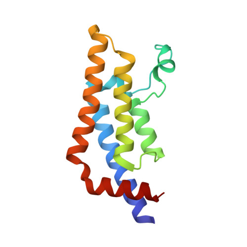
4TZ8

Structure of human ATAD2 bromodomain bound to fragment inhibitor


Experimental Data Snapshot

  • Method: X-RAY DIFFRACTION
  • Resolution: 2.15 Å
  • R-Value Free: 0.231 
  • R-Value Work: 0.198 
  • R-Value Observed: 0.202 

Starting Model: experimental
View more details

wwPDB Validation   3D Report Full Report


Ligand Structure Quality Assessment 


This is version 1.6 of the entry. See complete history


Literature

Fragment-Based Screening of the Bromodomain of ATAD2.

Harner, M.J.Chauder, B.A.Phan, J.Fesik, S.W.

(2014) J Med Chem 57: 9687-9692

  • DOI: https://doi.org/10.1021/jm501035j
  • Primary Citation of Related Structures:  
    4TYL, 4TZ2, 4TZ8

  • PubMed Abstract: 

    Cellular and genetic evidence suggest that inhibition of ATAD2 could be a useful strategy to treat several types of cancer. To discover small-molecule inhibitors of the bromodomain of ATAD2, we used a fragment-based approach. Fragment hits were identified using NMR spectroscopy, and ATAD2 was crystallized with three of the hits identified in the fragment screen.


  • Organizational Affiliation

    Department of Biochemistry, Vanderbilt University School of Medicine , 2215 Garland Avenue, 607 Light Hall, Nashville, Tennessee 37232-0146, United States.


Macromolecules
Find similar proteins by:  (by identity cutoff)  |  3D Structure
Entity ID: 1
MoleculeChains Sequence LengthOrganismDetailsImage
ATPase family AAA domain-containing protein 2130Homo sapiensMutation(s): 0 
Gene Names: ATAD2L16PRO2000
EC: 3.6.1.3
UniProt & NIH Common Fund Data Resources
Find proteins for Q6PL18 (Homo sapiens)
Explore Q6PL18 
Go to UniProtKB:  Q6PL18
PHAROS:  Q6PL18
GTEx:  ENSG00000156802 
Entity Groups  
Sequence Clusters30% Identity50% Identity70% Identity90% Identity95% Identity100% Identity
UniProt GroupQ6PL18
Sequence Annotations
Expand
  • Reference Sequence
Small Molecules
Binding Affinity Annotations 
IDSourceBinding Affinity
39U BindingDB:  4TZ8 Kd: 5.00e+5 (nM) from 1 assay(s)
Experimental Data & Validation

Experimental Data

  • Method: X-RAY DIFFRACTION
  • Resolution: 2.15 Å
  • R-Value Free: 0.231 
  • R-Value Work: 0.198 
  • R-Value Observed: 0.202 
  • Space Group: P 65 2 2
Unit Cell:
Length ( Å )Angle ( ˚ )
a = 79.942α = 90
b = 79.942β = 90
c = 138.259γ = 120
Software Package:
Software NamePurpose
PHENIXrefinement
HKL-2000data scaling
MAR345dtbdata collection
Cootmodel building

Structure Validation

View Full Validation Report


Ligand Structure Quality Assessment 


Entry History & Funding Information

Deposition Data


Funding OrganizationLocationGrant Number
National Institutes of Health/National Cancer Institute (NIH/NCI)United StatesDP1OD006933/DP1CA174419

Revision History  (Full details and data files)

  • Version 1.0: 2014-10-29
    Type: Initial release
  • Version 1.1: 2014-12-03
    Changes: Database references
  • Version 1.2: 2015-02-04
    Changes: Derived calculations
  • Version 1.3: 2017-09-06
    Changes: Author supporting evidence, Database references, Derived calculations, Other, Source and taxonomy
  • Version 1.4: 2017-11-22
    Changes: Refinement description
  • Version 1.5: 2019-12-04
    Changes: Author supporting evidence
  • Version 1.6: 2023-09-27
    Changes: Data collection, Database references, Refinement description