4TZ2 | pdb_00004tz2

Fragment-Based Screening of the Bromodomain of ATAD2


Experimental Data Snapshot

  • Method: X-RAY DIFFRACTION
  • Resolution: 1.70 Å
  • R-Value Free: 
    0.201 (Depositor), 0.225 (DCC) 
  • R-Value Work: 
    0.169 (Depositor) 
  • R-Value Observed: 
    0.171 (Depositor) 

Starting Model: experimental
View more details

wwPDB Validation 3D Report Full Report

Validation slider image for 4TZ2

Ligand Structure Quality Assessment 


This is version 1.8 of the entry. See complete history

Literature

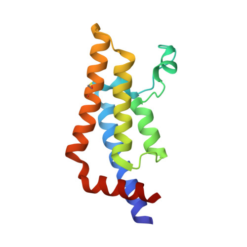
Fragment-Based Screening of the Bromodomain of ATAD2.

Harner, M.J.Chauder, B.A.Phan, J.Fesik, S.W.

(2014) J Med Chem 57: 9687-9692

  • DOI: https://doi.org/10.1021/jm501035j
  • Primary Citation Related Structures: 
    4TYL, 4TZ2, 4TZ8

  • PubMed Abstract: 

    Cellular and genetic evidence suggest that inhibition of ATAD2 could be a useful strategy to treat several types of cancer. To discover small-molecule inhibitors of the bromodomain of ATAD2, we used a fragment-based approach. Fragment hits were identified using NMR spectroscopy, and ATAD2 was crystallized with three of the hits identified in the fragment screen.


  • Organizational Affiliation
    • Department of Biochemistry, Vanderbilt University School of Medicine , 2215 Garland Avenue, 607 Light Hall, Nashville, Tennessee 37232-0146, United States.

Macromolecule Content 

  • Total Structure Weight: 15.99 kDa 
  • Atom Count: 1,363 
  • Modeled Residue Count: 130 
  • Deposited Residue Count: 130 
  • Unique protein chains: 1

Macromolecules

Find similar proteins by:|  3D Structure
Entity ID: 1
MoleculeChains  Sequence LengthOrganismDetailsImage
ATPase family AAA domain-containing protein 2130Homo sapiensMutation(s): 0 
Gene Names: ATAD2L16PRO2000
EC: 3.6.1.3 (PDB Primary Data), 3.6.1 (UniProt)
UniProt & NIH Common Fund Data Resources
Find proteins for Q6PL18 (Homo sapiens)
Explore Q6PL18 
Go to UniProtKB:  Q6PL18
PHAROS:  Q6PL18
GTEx:  ENSG00000156802 
Entity Groups
Sequence Clusters30% Identity50% Identity70% Identity90% Identity95% Identity100% Identity
UniProt GroupQ6PL18
Sequence Annotations
Expand
Reference Sequence

Small Molecules

Modified Residues  1 Unique
IDChains TypeFormula2D DiagramParent
CSO
Query on CSO
A
L-PEPTIDE LINKINGC3 H7 N O3 SCYS

Experimental Data & Validation

Experimental Data

  • Method: X-RAY DIFFRACTION
  • Resolution: 1.70 Å
  • R-Value Free:  0.201 (Depositor), 0.225 (DCC) 
  • R-Value Work:  0.169 (Depositor) 
  • R-Value Observed: 0.171 (Depositor) 
Space Group: P 65 2 2
Unit Cell:
Length ( Å )Angle ( ˚ )
a = 79.203α = 90
b = 79.203β = 90
c = 136.036γ = 120
Software Package:
Software NamePurpose
PHENIXrefinement
HKL-2000data scaling
MAR345dtbdata collection
Cootmodel building

Structure Validation

View Full Validation Report


Ligand Structure Quality Assessment 


Entry History 

& Funding Information

Deposition Data


Funding OrganizationLocationGrant Number
National Institutes of Health/National Cancer Institute (NIH/NCI)United StatesDP1OD006933/DP1CA174419

Revision History  (Full details and data files)

  • Version 1.0: 2014-10-29
    Type: Initial release
  • Version 1.1: 2014-12-03
    Changes: Database references
  • Version 1.2: 2015-02-04
    Changes: Derived calculations
  • Version 1.3: 2017-09-06
    Changes: Advisory, Author supporting evidence, Database references, Derived calculations, Other, Source and taxonomy
  • Version 1.4: 2017-11-22
    Changes: Refinement description
  • Version 1.5: 2019-12-04
    Changes: Author supporting evidence
  • Version 1.6: 2023-09-27
    Changes: Data collection, Database references, Refinement description
  • Version 1.7: 2023-11-15
    Changes: Data collection
  • Version 1.8: 2024-11-06
    Changes: Structure summary