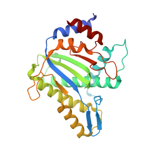
4R94

Structure of the nickase domain of NS1 from MVM complexed with magnesium


Experimental Data Snapshot

  • Method: X-RAY DIFFRACTION
  • Resolution: 1.67 Å
  • R-Value Free: 0.212 
  • R-Value Work: 0.175 
  • R-Value Observed: 0.177 

Starting Model: experimental
View more details

wwPDB Validation   3D Report Full Report


This is version 1.2 of the entry. See complete history


Literature

Structures of minute virus of mice replication initiator protein N-terminal domain: Insights into DNA nicking and origin binding.

Tewary, S.K.Liang, L.Lin, Z.Lynn, A.Cotmore, S.F.Tattersall, P.Zhao, H.Tang, L.

(2014) Virology 476C: 61-71

  • DOI: https://doi.org/10.1016/j.virol.2014.11.022
  • Primary Citation of Related Structures:  
    3WRN, 3WRO, 3WRQ, 3WRR, 3WRS, 4PP4, 4R94

  • PubMed Abstract: 

    Members of the Parvoviridae family all encode a non-structural protein 1 (NS1) that directs replication of single-stranded viral DNA, packages viral DNA into capsid, and serves as a potent transcriptional activator. Here we report the X-ray structure of the minute virus of mice (MVM) NS1 N-terminal domain at 1.45Å resolution, showing that sites for dsDNA binding, ssDNA binding and cleavage, nuclear localization, and other functions are integrated on a canonical fold of the histidine-hydrophobic-histidine superfamily of nucleases, including elements specific for this Protoparvovirus but distinct from its Bocaparvovirus or Dependoparvovirus orthologs. High resolution structural analysis reveals a nickase active site with an architecture that allows highly versatile metal ligand binding. The structures support a unified mechanism of replication origin recognition for homotelomeric and heterotelomeric parvoviruses, mediated by a basic-residue-rich hairpin and an adjacent helix in the initiator proteins and by tandem tetranucleotide motifs in the replication origins.


  • Organizational Affiliation

    Department of Molecular Biosciences, University of Kansas, Lawrence, KS 66045, USA.


Macromolecules
Find similar proteins by:  (by identity cutoff)  |  3D Structure
Entity ID: 1
MoleculeChains Sequence LengthOrganismDetailsImage
Non-structural protein NS1255Murine minute virus strain MVM prototypeMutation(s): 0 
Gene Names: NS1
EC: 3.1.21 (UniProt), 3.6.4.12 (UniProt)
UniProt
Find proteins for P03134 (Murine minute virus (strain MVM prototype))
Explore P03134 
Go to UniProtKB:  P03134
Entity Groups  
Sequence Clusters30% Identity50% Identity70% Identity90% Identity95% Identity100% Identity
UniProt GroupP03134
Sequence Annotations
Expand
  • Reference Sequence
Small Molecules
Ligands 1 Unique
IDChains Name / Formula / InChI Key2D Diagram3D Interactions
MG
Query on MG

Download Ideal Coordinates CCD File 
B [auth A]MAGNESIUM ION
Mg
JLVVSXFLKOJNIY-UHFFFAOYSA-N
Experimental Data & Validation

Experimental Data

  • Method: X-RAY DIFFRACTION
  • Resolution: 1.67 Å
  • R-Value Free: 0.212 
  • R-Value Work: 0.175 
  • R-Value Observed: 0.177 
  • Space Group: P 21 21 2
Unit Cell:
Length ( Å )Angle ( ˚ )
a = 57.016α = 90
b = 121.377β = 90
c = 41.581γ = 90
Software Package:
Software NamePurpose
Blu-Icedata collection
PHENIXmodel building
PHENIXrefinement
HKL-2000data reduction
HKL-2000data scaling
PHENIXphasing

Structure Validation

View Full Validation Report


Entry History 

Deposition Data

Revision History  (Full details and data files)

  • Version 1.0: 2015-01-21
    Type: Initial release
  • Version 1.1: 2017-11-22
    Changes: Refinement description
  • Version 1.2: 2023-11-08
    Changes: Data collection, Database references, Derived calculations, Refinement description