4PP4 | pdb_00004pp4

Minute virus of mice non-structural protein-1N-terminal nuclease domain reveals a unique Zn2+ coordination in the active site pocket and shows a novel mode of DNA recognition at the origin of replication


Experimental Data Snapshot

  • Method: X-RAY DIFFRACTION
  • Resolution: 1.45 Å
  • R-Value Free: 
    0.185 (Depositor), 0.190 (DCC) 
  • R-Value Work: 
    0.166 (Depositor), 0.169 (DCC) 
  • R-Value Observed: 
    0.167 (Depositor) 

wwPDB Validation 3D Report Full Report

Validation slider image for 4PP4

This is version 1.1 of the entry. See complete history

Literature

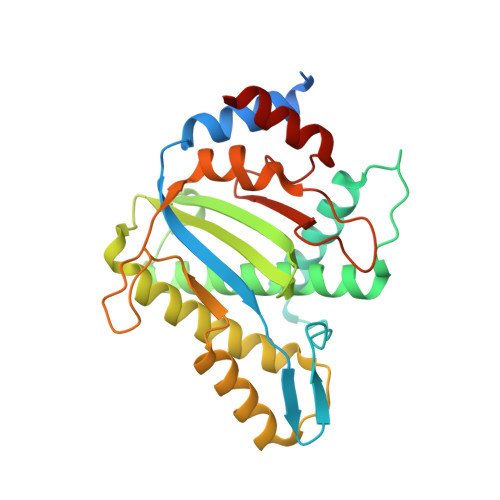
Structures of minute virus of mice replication initiator protein N-terminal domain: Insights into DNA nicking and origin binding.

Tewary, S.K.Liang, L.Lin, Z.Lynn, A.Cotmore, S.F.Tattersall, P.Zhao, H.Tang, L.

(2014) Virology 476C: 61-71

  • DOI: https://doi.org/10.1016/j.virol.2014.11.022
  • Primary Citation Related Structures: 
    3WRN, 3WRO, 3WRQ, 3WRR, 3WRS, 4PP4, 4R94

  • PubMed Abstract: 

    Members of the Parvoviridae family all encode a non-structural protein 1 (NS1) that directs replication of single-stranded viral DNA, packages viral DNA into capsid, and serves as a potent transcriptional activator. Here we report the X-ray structure of the minute virus of mice (MVM) NS1 N-terminal domain at 1.45Å resolution, showing that sites for dsDNA binding, ssDNA binding and cleavage, nuclear localization, and other functions are integrated on a canonical fold of the histidine-hydrophobic-histidine superfamily of nucleases, including elements specific for this Protoparvovirus but distinct from its Bocaparvovirus or Dependoparvovirus orthologs. High resolution structural analysis reveals a nickase active site with an architecture that allows highly versatile metal ligand binding. The structures support a unified mechanism of replication origin recognition for homotelomeric and heterotelomeric parvoviruses, mediated by a basic-residue-rich hairpin and an adjacent helix in the initiator proteins and by tandem tetranucleotide motifs in the replication origins.


  • Organizational Affiliation
    • Department of Molecular Biosciences, University of Kansas, Lawrence, KS 66045, USA.

Macromolecule Content 

  • Total Structure Weight: 32.49 kDa 
  • Atom Count: 2,323 
  • Modeled Residue Count: 248 
  • Deposited Residue Count: 276 
  • Unique protein chains: 1

Macromolecules

Find similar proteins by:|  3D Structure
Entity ID: 1
MoleculeChains  Sequence LengthOrganismDetailsImage
Non-capsid protein NS-1276Murine minute virus strain MVM prototypeMutation(s): 0 
Gene Names: NS1
EC: 3.1.21 (UniProt), 3.6.4.12 (UniProt)
UniProt
Find proteins for P03134 (Murine minute virus (strain MVM prototype))
Explore P03134 
Go to UniProtKB:  P03134
Entity Groups
Sequence Clusters30% Identity50% Identity70% Identity90% Identity95% Identity100% Identity
UniProt GroupP03134
Sequence Annotations
Expand
Reference Sequence

Experimental Data & Validation

Experimental Data

  • Method: X-RAY DIFFRACTION
  • Resolution: 1.45 Å
  • R-Value Free:  0.185 (Depositor), 0.190 (DCC) 
  • R-Value Work:  0.166 (Depositor), 0.169 (DCC) 
  • R-Value Observed: 0.167 (Depositor) 
Space Group: P 21 21 2
Unit Cell:
Length ( Å )Angle ( ˚ )
a = 56.94α = 90
b = 121.65β = 90
c = 41.82γ = 90
Software Package:
Software NamePurpose
SOLVEphasing
PHENIXrefinement
XDSdata reduction
SCALAdata scaling

Structure Validation

View Full Validation Report


Entry History 

Deposition Data

Revision History  (Full details and data files)

  • Version 1.0: 2015-01-21
    Type: Initial release
  • Version 1.1: 2024-03-20
    Changes: Data collection, Database references, Derived calculations