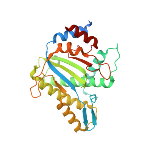
Structures of minute virus of mice replication initiator protein N-terminal domain: Insights into DNA nicking and origin binding.
Tewary, S.K., Liang, L., Lin, Z., Lynn, A., Cotmore, S.F., Tattersall, P., Zhao, H., Tang, L.(2014) Virology 476C: 61-71
- PubMed: 25528417
- DOI: https://doi.org/10.1016/j.virol.2014.11.022
- Primary Citation of Related Structures:
3WRN, 3WRO, 3WRQ, 3WRR, 3WRS, 4PP4, 4R94 - PubMed Abstract:
Members of the Parvoviridae family all encode a non-structural protein 1 (NS1) that directs replication of single-stranded viral DNA, packages viral DNA into capsid, and serves as a potent transcriptional activator. Here we report the X-ray structure of the minute virus of mice (MVM) NS1 N-terminal domain at 1.45Å resolution, showing that sites for dsDNA binding, ssDNA binding and cleavage, nuclear localization, and other functions are integrated on a canonical fold of the histidine-hydrophobic-histidine superfamily of nucleases, including elements specific for this Protoparvovirus but distinct from its Bocaparvovirus or Dependoparvovirus orthologs. High resolution structural analysis reveals a nickase active site with an architecture that allows highly versatile metal ligand binding. The structures support a unified mechanism of replication origin recognition for homotelomeric and heterotelomeric parvoviruses, mediated by a basic-residue-rich hairpin and an adjacent helix in the initiator proteins and by tandem tetranucleotide motifs in the replication origins.
- Department of Molecular Biosciences, University of Kansas, Lawrence, KS 66045, USA.
Organizational Affiliation:


