4R2A | pdb_00004r2a

Egr1/Zif268 zinc fingers in complex with methylated DNA


Experimental Data Snapshot

  • Method: X-RAY DIFFRACTION
  • Resolution: 1.59 Å
  • R-Value Free: 
    0.189 (Depositor), 0.195 (DCC) 
  • R-Value Work: 
    0.137 (Depositor), 0.148 (DCC) 
  • R-Value Observed: 
    0.140 (Depositor) 

Starting Model: experimental
View more details

wwPDB Validation 3D Report Full Report

Validation slider image for 4R2A

This is version 1.2 of the entry. See complete history

Literature

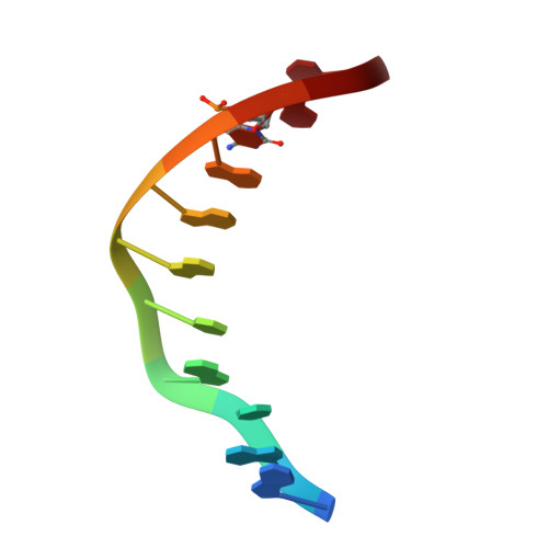
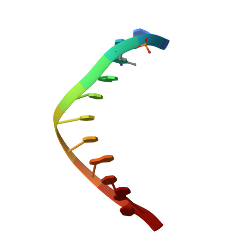
Wilms tumor protein recognizes 5-carboxylcytosine within a specific DNA sequence.

Hashimoto, H.Olanrewaju, Y.O.Zheng, Y.Wilson, G.G.Zhang, X.Cheng, X.

(2014) Genes Dev 28: 2304-2313

  • DOI: https://doi.org/10.1101/gad.250746.114
  • Primary Citation Related Structures: 
    4R2A, 4R2C, 4R2D, 4R2E, 4R2P, 4R2Q, 4R2R, 4R2S

  • PubMed Abstract: 

    In mammalian DNA, cytosine occurs in several chemical forms, including unmodified cytosine (C), 5-methylcytosine (5 mC), 5-hydroxymethylcytosine (5 hmC), 5-formylcytosine (5 fC), and 5-carboxylcytosine (5 caC). 5 mC is a major epigenetic signal that acts to regulate gene expression. 5 hmC, 5 fC, and 5 caC are oxidized derivatives that might also act as distinct epigenetic signals. We investigated the response of the zinc finger DNA-binding domains of transcription factors early growth response protein 1 (Egr1) and Wilms tumor protein 1 (WT1) to different forms of modified cytosine within their recognition sequence, 5'-GCG(T/G)GGGCG-3'. Both displayed high affinity for the sequence when C or 5 mC was present and much reduced affinity when 5 hmC or 5 fC was present, indicating that they differentiate primarily oxidized C from unoxidized C, rather than methylated C from unmethylated C. 5 caC affected the two proteins differently, abolishing binding by Egr1 but not by WT1. We ascribe this difference to electrostatic interactions in the binding sites. In Egr1, a negatively charged glutamate conflicts with the negatively charged carboxylate of 5 caC, whereas the corresponding glutamine of WT1 interacts with this group favorably. Our analyses shows that zinc finger proteins (and their splice variants) can respond in modulated ways to alternative modifications within their binding sequence.


  • Organizational Affiliation
    • Department of Biochemistry, Emory University School of Medicine, Atlanta, Georgia 30322, USA;

Macromolecule Content 

  • Total Structure Weight: 18.19 kDa 
  • Atom Count: 1,413 
  • Modeled Residue Count: 108 
  • Deposited Residue Count: 116 
  • Unique protein chains: 1
  • Unique nucleic acid chains: 2

Macromolecules


Find similar proteins by:|  3D Structure
Entity ID: 1
MoleculeChains  Sequence LengthOrganismDetailsImage
Early growth response protein 194Homo sapiensMutation(s): 0 
Gene Names: EGR1KROX24ZNF225
UniProt & NIH Common Fund Data Resources
Find proteins for P18146 (Homo sapiens)
Explore P18146 
Go to UniProtKB:  P18146
PHAROS:  P18146
GTEx:  ENSG00000120738 
Entity Groups
Sequence Clusters30% Identity50% Identity70% Identity90% Identity95% Identity100% Identity
UniProt GroupP18146
Sequence Annotations
Expand
Reference Sequence
Find similar nucleic acids by:  Sequence
Entity ID: 2
MoleculeChains LengthOrganismImage
DNA (5'-D(*AP*GP*CP*GP*TP*GP*GP*GP*(5CM)P*GP*T)-3')11synthetic construct
Sequence Annotations
Expand
Reference Sequence
Find similar nucleic acids by:  Sequence
Entity ID: 3
MoleculeChains LengthOrganismImage
DNA (5'-D(*TP*AP*(5CM)P*GP*CP*CP*CP*AP*CP*GP*C)-3')11synthetic construct
Sequence Annotations
Expand
Reference Sequence

Experimental Data & Validation

Experimental Data

  • Method: X-RAY DIFFRACTION
  • Resolution: 1.59 Å
  • R-Value Free:  0.189 (Depositor), 0.195 (DCC) 
  • R-Value Work:  0.137 (Depositor), 0.148 (DCC) 
  • R-Value Observed: 0.140 (Depositor) 
Space Group: C 2 2 21
Unit Cell:
Length ( Å )Angle ( ˚ )
a = 43.99α = 90
b = 56.208β = 90
c = 130.219γ = 90
Software Package:
Software NamePurpose
HKL-2000data collection
PHENIXmodel building
PHENIXrefinement
HKL-2000data reduction
HKL-2000data scaling
PHENIXphasing

Structure Validation

View Full Validation Report


Entry History 

Deposition Data

Revision History  (Full details and data files)

  • Version 1.0: 2014-10-08
    Type: Initial release
  • Version 1.1: 2014-11-19
    Changes: Database references
  • Version 1.2: 2023-09-20
    Changes: Data collection, Database references, Derived calculations, Refinement description