4Q7J | pdb_00004q7j

Complex structure of viral RNA polymerase


Experimental Data Snapshot

  • Method: X-RAY DIFFRACTION
  • Resolution: 2.90 Å
  • R-Value Free: 
    0.309 (Depositor), 0.303 (DCC) 
  • R-Value Work: 
    0.255 (Depositor), 0.254 (DCC) 
  • R-Value Observed: 
    0.258 (Depositor) 

Starting Model: experimental
View more details

wwPDB Validation 3D Report Full Report

Validation slider image for 4Q7J

This is version 1.2 of the entry. See complete history

Literature

Molecular insights into replication initiation by Q beta replicase using ribosomal protein S1.

Takeshita, D.Yamashita, S.Tomita, K.

(2014) Nucleic Acids Res 42: 10809-10822

  • DOI: https://doi.org/10.1093/nar/gku745
  • Primary Citation Related Structures: 
    4Q7J

  • PubMed Abstract: 

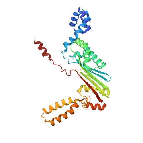
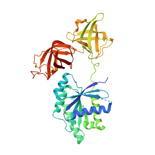
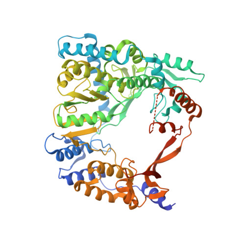
    Ribosomal protein S1, consisting of six contiguous OB-folds, is the largest ribosomal protein and is essential for translation initiation in Escherichia coli. S1 is also one of the three essential host-derived subunits of Qβ replicase, together with EF-Tu and EF-Ts, for Qβ RNA replication in E. coli. We analyzed the crystal structure of Qβ replicase, consisting of the virus-encoded RNA-dependent RNA polymerase (β-subunit), EF-Tu, EF-Ts and the N-terminal half of S1, which is capable of initiating Qβ RNA replication. Structural and biochemical studies revealed that the two N-terminal OB-folds of S1 anchor S1 onto the β-subunit, and the third OB-fold is mobile and protrudes beyond the surface of the β-subunit. The third OB-fold mainly interacts with a specific RNA fragment derived from the internal region of Qβ RNA, and its RNA-binding ability is required for replication initiation of Qβ RNA. Thus, the third mobile OB-fold of S1, which is spatially anchored near the surface of the β-subunit, primarily recruits the Qβ RNA toward the β-subunit, leading to the specific and efficient replication initiation of Qβ RNA, and S1 functions as a replication initiation factor, beyond its established function in protein synthesis.


  • Organizational Affiliation
    • Biomedical Research Institute, National Institute of Advanced Industrial Science and Technology (AIST), Tsukuba, Ibaraki 305-8566, Japan.

Macromolecule Content 

  • Total Structure Weight: 342.76 kDa 
  • Atom Count: 21,369 
  • Modeled Residue Count: 2,730 
  • Deposited Residue Count: 3,100 
  • Unique protein chains: 4

Macromolecules

Find similar proteins by:|  3D Structure
Entity ID: 1
MoleculeChains  Sequence LengthOrganismDetailsImage
Elongation factor Ts
A, E
282Escherichia coli K-12Mutation(s): 0 
Gene Names: tsf
UniProt
Find proteins for P0A6P1 (Escherichia coli (strain K12))
Explore P0A6P1 
Go to UniProtKB:  P0A6P1
Entity Groups
Sequence Clusters30% Identity50% Identity70% Identity90% Identity95% Identity100% Identity
UniProt GroupP0A6P1
Sequence Annotations
Expand
Reference Sequence
Find similar proteins by:|  3D Structure
Entity ID: 2
MoleculeChains  Sequence LengthOrganismDetailsImage
Elongation factor Tu 1
B, F
393Escherichia coli K-12Mutation(s): 0 
Gene Names: tufA
EC: 3.6.5.3
UniProt
Find proteins for P0CE47 (Escherichia coli (strain K12))
Explore P0CE47 
Go to UniProtKB:  P0CE47
Entity Groups
Sequence Clusters30% Identity50% Identity70% Identity90% Identity95% Identity100% Identity
UniProt GroupP0CE47
Sequence Annotations
Expand
Reference Sequence
Find similar proteins by:|  3D Structure
Entity ID: 3
MoleculeChains  Sequence LengthOrganismDetailsImage
Q beta replicase
C, G
594Qubevirus durumMutation(s): 0 
EC: 2.7.7.48
UniProt
Find proteins for P14647 (Escherichia virus Qbeta)
Explore P14647 
Go to UniProtKB:  P14647
Entity Groups
Sequence Clusters30% Identity50% Identity70% Identity90% Identity95% Identity100% Identity
UniProt GroupP14647
Sequence Annotations
Expand
Reference Sequence
Find similar proteins by:|  3D Structure
Entity ID: 4
MoleculeChains  Sequence LengthOrganismDetailsImage
30S ribosomal protein S1
D, H
281Escherichia coli K-12Mutation(s): 0 
Gene Names: rpsA
UniProt
Find proteins for P0AG67 (Escherichia coli (strain K12))
Explore P0AG67 
Go to UniProtKB:  P0AG67
Entity Groups
Sequence Clusters30% Identity50% Identity70% Identity90% Identity95% Identity100% Identity
UniProt GroupP0AG67
Sequence Annotations
Expand
Reference Sequence

Experimental Data & Validation

Experimental Data

  • Method: X-RAY DIFFRACTION
  • Resolution: 2.90 Å
  • R-Value Free:  0.309 (Depositor), 0.303 (DCC) 
  • R-Value Work:  0.255 (Depositor), 0.254 (DCC) 
  • R-Value Observed: 0.258 (Depositor) 
Space Group: P 21 21 21
Unit Cell:
Length ( Å )Angle ( ˚ )
a = 132.19α = 90
b = 150.83β = 90
c = 189.78γ = 90
Software Package:
Software NamePurpose
HKL-2000data collection
PHASERphasing
PHENIXrefinement
HKL-2000data reduction
HKL-2000data scaling

Structure Validation

View Full Validation Report


Entry History 

Deposition Data

Revision History  (Full details and data files)

  • Version 1.0: 2014-08-27
    Type: Initial release
  • Version 1.1: 2022-08-24
    Changes: Database references, Derived calculations
  • Version 1.2: 2023-11-08
    Changes: Data collection, Refinement description