4LNI

B. subtilis glutamine synthetase structures reveal large active site conformational changes and basis for isoenzyme specific regulation: structure of the transition state complex


Experimental Data Snapshot

  • Method: X-RAY DIFFRACTION
  • Resolution: 2.58 Å
  • R-Value Free: 0.223 
  • R-Value Work: 0.165 
  • R-Value Observed: 0.166 

wwPDB Validation   3D Report Full Report


Ligand Structure Quality Assessment 


This is version 1.4 of the entry. See complete history


Literature

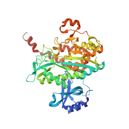
Structures of the Bacillus subtilis Glutamine Synthetase Dodecamer Reveal Large Intersubunit Catalytic Conformational Changes Linked to a Unique Feedback Inhibition Mechanism.

Murray, D.S.Chinnam, N.Tonthat, N.K.Whitfill, T.Wray, L.V.Fisher, S.H.Schumacher, M.A.

(2013) J Biol Chem 288: 35801-35811

  • DOI: https://doi.org/10.1074/jbc.M113.519496
  • Primary Citation of Related Structures:  
    4LNF, 4LNI, 4LNK, 4LNN, 4LNO

  • PubMed Abstract: 

    Glutamine synthetase (GS), which catalyzes the production of glutamine, plays essential roles in nitrogen metabolism. There are two main bacterial GS isoenzymes, GSI-α and GSI-β. GSI-α enzymes, which have not been structurally characterized, are uniquely feedback-inhibited by Gln. To gain insight into GSI-α function, we performed biochemical and cellular studies and obtained structures for all GSI-α catalytic and regulatory states. GSI-α forms a massive 600-kDa dodecameric machine. Unlike other characterized GS, the Bacillus subtilis enzyme undergoes dramatic intersubunit conformational alterations during formation of the transition state. Remarkably, these changes are required for active site construction. Feedback inhibition arises from a hydrogen bond network between Gln, the catalytic glutamate, and the GSI-α-specific residue, Arg(62), from an adjacent subunit. Notably, Arg(62) must be ejected for proper active site reorganization. Consistent with these findings, an R62A mutation abrogates Gln feedback inhibition but does not affect catalysis. Thus, these data reveal a heretofore unseen restructuring of an enzyme active site that is coupled with an isoenzyme-specific regulatory mechanism. This GSI-α-specific regulatory network could be exploited for inhibitor design against Gram-positive pathogens.


  • Organizational Affiliation

    From the Department of Biochemistry and Molecular Biology, Oregon Health and Science University, Portland, Oregon 97239.


Macromolecules
Find similar proteins by:  (by identity cutoff)  |  3D Structure
Entity ID: 1
MoleculeChains Sequence LengthOrganismDetailsImage
Glutamine synthetase
A, B, C, D, E
A, B, C, D, E, F, G, H, I, J, K, L
443Bacillus subtilisMutation(s): 0 
Gene Names: glnABSU17460
EC: 6.3.1.2
UniProt
Find proteins for P12425 (Bacillus subtilis (strain 168))
Explore P12425 
Go to UniProtKB:  P12425
Entity Groups  
Sequence Clusters30% Identity50% Identity70% Identity90% Identity95% Identity100% Identity
UniProt GroupP12425
Sequence Annotations
Expand
  • Reference Sequence
Small Molecules
Ligands 3 Unique
IDChains Name / Formula / InChI Key2D Diagram3D Interactions
ADP
Query on ADP

Download Ideal Coordinates CCD File 
BB [auth I]
CA [auth D]
GB [auth J]
HA [auth E]
LB [auth K]
BB [auth I],
CA [auth D],
GB [auth J],
HA [auth E],
LB [auth K],
MA [auth F],
N [auth A],
QB [auth L],
RA [auth G],
S [auth B],
WA [auth H],
X [auth C]
ADENOSINE-5'-DIPHOSPHATE
C10 H15 N5 O10 P2
XTWYTFMLZFPYCI-KQYNXXCUSA-N
P3S
Query on P3S

Download Ideal Coordinates CCD File 
AB [auth I]
BA [auth D]
FB [auth J]
GA [auth E]
KB [auth K]
AB [auth I],
BA [auth D],
FB [auth J],
GA [auth E],
KB [auth K],
LA [auth F],
M [auth A],
PB [auth L],
QA [auth G],
R [auth B],
VA [auth H],
W [auth C]
L-METHIONINE-S-SULFOXIMINE PHOSPHATE
C5 H13 N2 O6 P S
QQFOFBSCSWFFPB-NMAPHRJESA-N
MG
Query on MG

Download Ideal Coordinates CCD File 
AA [auth C]
CB [auth I]
DA [auth D]
DB [auth I]
EA [auth D]
AA [auth C],
CB [auth I],
DA [auth D],
DB [auth I],
EA [auth D],
EB [auth I],
FA [auth D],
HB [auth J],
IA [auth E],
IB [auth J],
JA [auth E],
JB [auth J],
KA [auth E],
MB [auth K],
NA [auth F],
NB [auth K],
O [auth A],
OA [auth F],
OB [auth K],
P [auth A],
PA [auth F],
Q [auth A],
RB [auth L],
SA [auth G],
SB [auth L],
T [auth B],
TA [auth G],
TB [auth L],
U [auth B],
UA [auth G],
V [auth B],
XA [auth H],
Y [auth C],
YA [auth H],
Z [auth C],
ZA [auth H]
MAGNESIUM ION
Mg
JLVVSXFLKOJNIY-UHFFFAOYSA-N
Experimental Data & Validation

Experimental Data

  • Method: X-RAY DIFFRACTION
  • Resolution: 2.58 Å
  • R-Value Free: 0.223 
  • R-Value Work: 0.165 
  • R-Value Observed: 0.166 
  • Space Group: P 1
Unit Cell:
Length ( Å )Angle ( ˚ )
a = 110.2α = 60.29
b = 141.6β = 67.38
c = 142.1γ = 76.2
Software Package:
Software NamePurpose
PHENIXrefinement
MOLREPphasing
CNSrefinement
MOSFLMdata reduction
SCALAdata scaling
ADSCdata collection

Structure Validation

View Full Validation Report


Ligand Structure Quality Assessment 


Entry History 

Deposition Data

Revision History  (Full details and data files)

  • Version 1.0: 2013-11-06
    Type: Initial release
  • Version 1.1: 2013-11-13
    Changes: Database references
  • Version 1.2: 2014-01-15
    Changes: Database references
  • Version 1.3: 2019-07-17
    Changes: Data collection, Refinement description
  • Version 1.4: 2024-02-28
    Changes: Data collection, Database references, Derived calculations