4L79 | pdb_00004l79

Crystal Structure of nucleotide-free Myosin 1b residues 1-728 with bound Calmodulin


Experimental Data Snapshot

  • Method: X-RAY DIFFRACTION
  • Resolution: 2.30 Å
  • R-Value Free: 
    0.249 (Depositor), 0.251 (DCC) 
  • R-Value Work: 
    0.189 (Depositor), 0.191 (DCC) 
  • R-Value Observed: 
    0.192 (Depositor) 

Starting Model: experimental
View more details

wwPDB Validation 3D Report Full Report

Validation slider image for 4L79

This is version 1.2 of the entry. See complete history

Literature

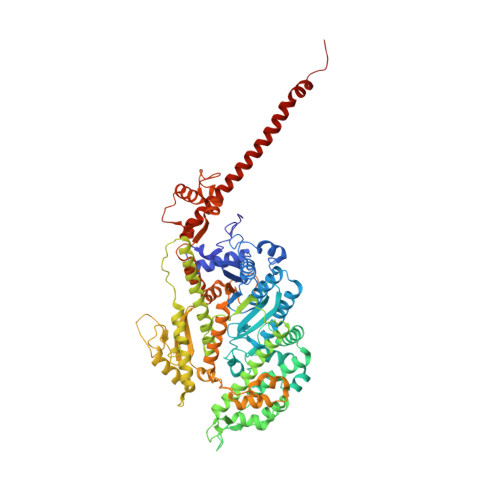
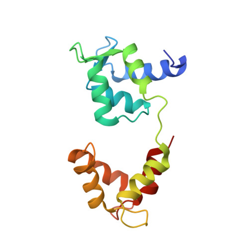
A vertebrate myosin-I structure reveals unique insights into myosin mechanochemical tuning.

Shuman, H.Greenberg, M.J.Zwolak, A.Lin, T.Sindelar, C.V.Dominguez, R.Ostap, E.M.

(2014) Proc Natl Acad Sci U S A 111: 2116-2121

  • DOI: https://doi.org/10.1073/pnas.1321022111
  • Primary Citation Related Structures: 
    4L79

  • PubMed Abstract: 

    Myosins are molecular motors that power diverse cellular processes, such as rapid organelle transport, muscle contraction, and tension-sensitive anchoring. The structural adaptations in the motor that allow for this functional diversity are not known, due, in part, to the lack of high-resolution structures of highly tension-sensitive myosins. We determined a 2.3-Å resolution structure of apo-myosin-Ib (Myo1b), which is the most tension-sensitive myosin characterized. We identified a striking unique orientation of structural elements that position the motor's lever arm. This orientation results in a cavity between the motor and lever arm that holds a 10-residue stretch of N-terminal amino acids, a region that is divergent among myosins. Single-molecule and biochemical analyses show that the N terminus plays an important role in stabilizing the post power-stroke conformation of Myo1b and in tuning the rate of the force-sensitive transition. We propose that this region plays a general role in tuning the mechanochemical properties of myosins.


  • Organizational Affiliation
    • Pennsylvania Muscle Institute and Department of Physiology, Perelman School of Medicine at the University of Pennsylvania, Philadelphia, PA 19104.

Macromolecule Content 

  • Total Structure Weight: 102.88 kDa 
  • Atom Count: 7,631 
  • Modeled Residue Count: 881 
  • Deposited Residue Count: 893 
  • Unique protein chains: 2

Macromolecules

Find similar proteins by:|  3D Structure
Entity ID: 1
MoleculeChains  Sequence LengthOrganismDetailsImage
Unconventional myosin-Ib744Rattus norvegicusMutation(s): 0 
Gene Names: Myo1aMyo1bMyr1
UniProt
Find proteins for Q05096 (Rattus norvegicus)
Explore Q05096 
Go to UniProtKB:  Q05096
Entity Groups
Sequence Clusters30% Identity50% Identity70% Identity90% Identity95% Identity100% Identity
UniProt GroupQ05096
Sequence Annotations
Expand
Reference Sequence
Find similar proteins by:|  3D Structure
Entity ID: 2
MoleculeChains  Sequence LengthOrganismDetailsImage
Calmodulin149Homo sapiensMutation(s): 0 
Gene Names: 
UniProt & NIH Common Fund Data Resources
Find proteins for P0DP23 (Homo sapiens)
Explore P0DP23 
Go to UniProtKB:  P0DP23
PHAROS:  P0DP23
GTEx:  ENSG00000198668 
Entity Groups
Sequence Clusters30% Identity50% Identity70% Identity90% Identity95% Identity100% Identity
UniProt GroupP0DP23
Sequence Annotations
Expand
Reference Sequence

Small Molecules

Ligands 1 Unique
IDChains Name / Formula / InChI Key2D Diagram3D Interactions
MG

Query on MG


Download:Ideal Coordinates CCD File
C [auth A]MAGNESIUM ION
Mg
JLVVSXFLKOJNIY-UHFFFAOYSA-N

Experimental Data & Validation

Experimental Data

  • Method: X-RAY DIFFRACTION
  • Resolution: 2.30 Å
  • R-Value Free:  0.249 (Depositor), 0.251 (DCC) 
  • R-Value Work:  0.189 (Depositor), 0.191 (DCC) 
  • R-Value Observed: 0.192 (Depositor) 
Space Group: P 1 21 1
Unit Cell:
Length ( Å )Angle ( ˚ )
a = 114.184α = 90
b = 45.09β = 117.18
c = 115.547γ = 90
Software Package:
Software NamePurpose
ADSCdata collection
PHENIXmodel building
PHENIXrefinement
HKL-2000data reduction
HKL-2000data scaling
PHENIXphasing

Structure Validation

View Full Validation Report


Entry History 

Deposition Data

Revision History  (Full details and data files)

  • Version 1.0: 2014-01-29
    Type: Initial release
  • Version 1.1: 2014-02-26
    Changes: Database references
  • Version 1.2: 2023-09-20
    Changes: Data collection, Database references, Derived calculations, Refinement description