4K9E | pdb_00004k9e

Crystal structure of KIT D4D5 fragment in complex with anti-Kit antibodies Fab79D


Experimental Data Snapshot

  • Method: X-RAY DIFFRACTION
  • Resolution: 2.70 Å
  • R-Value Free: 
    0.282 (Depositor), 0.283 (DCC) 
  • R-Value Work: 
    0.247 (Depositor), 0.247 (DCC) 
  • R-Value Observed: 
    0.248 (Depositor) 

wwPDB Validation 3D Report Full Report

Validation slider image for 4K9E

Ligand Structure Quality Assessment 


This is version 1.3 of the entry. See complete history

Literature

Structural basis for KIT receptor tyrosine kinase inhibition by antibodies targeting the D4 membrane-proximal region.

Reshetnyak, A.V.Nelson, B.Shi, X.Boggon, T.J.Pavlenco, A.Mandel-Bausch, E.M.Tome, F.Suzuki, Y.Sidhu, S.S.Lax, I.Schlessinger, J.

(2013) Proc Natl Acad Sci U S A 110: 17832-17837

  • DOI: https://doi.org/10.1073/pnas.1317118110
  • Primary Citation Related Structures: 
    4K94, 4K9E

  • PubMed Abstract: 

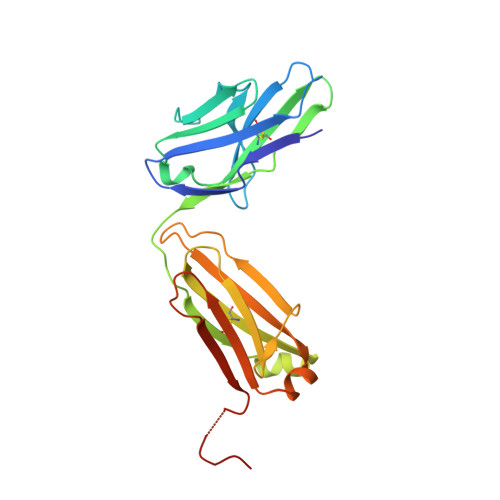
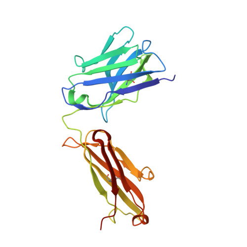
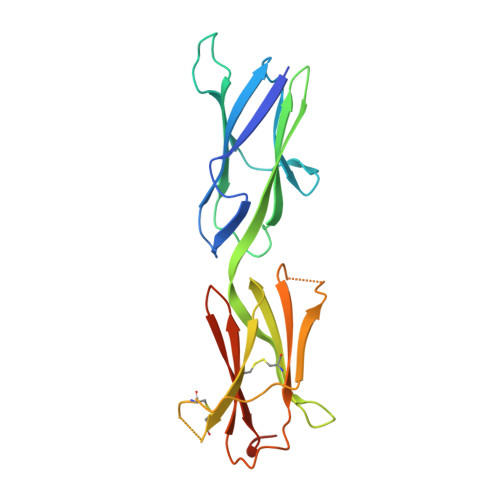
    Somatic oncogenic mutations in the receptor tyrosine kinase KIT function as major drivers of gastrointestinal stromal tumors and a subset of acute myeloid leukemia, melanoma, and other cancers. Although treatment of these cancers with tyrosine kinase inhibitors shows dramatic responses and durable disease control, drug resistance followed by clinical progression of disease eventually occurs in virtually all patients. In this report, we describe inhibitory KIT antibodies that bind to the membrane-proximal Ig-like D4 of KIT with significant overlap with an epitope in D4 that mediates homotypic interactions essential for KIT activation. Crystal structures of the anti-KIT antibody in complex with KIT D4 and D5 allowed design of affinity-matured libraries that were used to isolate variants with increased affinity and efficacy. Isolated antibodies showed KIT inhibition together with suppression of cell proliferation driven by ligand-stimulated WT or constitutively activated oncogenic KIT mutant. These antibodies represent a unique therapeutic approach and a step toward the development of "naked" or toxin-conjugated KIT antibodies for the treatment of KIT-driven cancers.


  • Organizational Affiliation
    • Department of Pharmacology, Yale University School of Medicine, New Haven, CT 06520.

Macromolecule Content 

  • Total Structure Weight: 72.75 kDa 
  • Atom Count: 4,949 
  • Modeled Residue Count: 634 
  • Deposited Residue Count: 663 
  • Unique protein chains: 3

Macromolecules

Find similar proteins by:|  3D Structure
Entity ID: 1
MoleculeChains  Sequence LengthOrganismDetailsImage
light chainA [auth L]228Mus musculusMutation(s): 0 
Find similar proteins by:|  3D Structure
Entity ID: 2
MoleculeChains  Sequence LengthOrganismDetailsImage
heavy chainB [auth H]221Mus musculusMutation(s): 0 
Find similar proteins by:|  3D Structure
Entity ID: 3
MoleculeChains  Sequence LengthOrganismDetailsImage
Mast/stem cell growth factor receptor Kit214Homo sapiensMutation(s): 0 
Gene Names: KITSCFR
EC: 2.7.10.1
UniProt & NIH Common Fund Data Resources
Find proteins for P10721 (Homo sapiens)
Explore P10721 
Go to UniProtKB:  P10721
PHAROS:  P10721
GTEx:  ENSG00000157404 
Entity Groups
Sequence Clusters30% Identity50% Identity70% Identity90% Identity95% Identity100% Identity
UniProt GroupP10721
Glycosylation
Glycosylation Sites: 1Go to GlyGen: P10721-1
Sequence Annotations
Expand
Reference Sequence

Small Molecules

Ligands 1 Unique
IDChains Name / Formula / InChI Key2D Diagram3D Interactions
NAG

Query on NAG


Download:Ideal Coordinates CCD File
D [auth C]2-acetamido-2-deoxy-beta-D-glucopyranose
C8 H15 N O6
OVRNDRQMDRJTHS-FMDGEEDCSA-N

Experimental Data & Validation

Experimental Data

  • Method: X-RAY DIFFRACTION
  • Resolution: 2.70 Å
  • R-Value Free:  0.282 (Depositor), 0.283 (DCC) 
  • R-Value Work:  0.247 (Depositor), 0.247 (DCC) 
  • R-Value Observed: 0.248 (Depositor) 
Space Group: P 65 2 2
Unit Cell:
Length ( Å )Angle ( ˚ )
a = 94.577α = 90
b = 94.577β = 90
c = 320.876γ = 120
Software Package:
Software NamePurpose
DENZOdata reduction
SCALEPACKdata scaling
PHASERphasing
PHENIXrefinement
PDB_EXTRACTdata extraction
CBASSdata collection

Structure Validation

View Full Validation Report


Ligand Structure Quality Assessment 


Entry History 

Deposition Data

Revision History  (Full details and data files)

  • Version 1.0: 2013-10-16
    Type: Initial release
  • Version 1.1: 2014-03-12
    Changes: Database references
  • Version 1.2: 2020-07-29
    Type: Remediation
    Reason: Carbohydrate remediation
    Changes: Data collection, Database references, Derived calculations, Structure summary
  • Version 1.3: 2024-11-27
    Changes: Data collection, Database references, Structure summary