Structural and Biophysical Characterization of the Cytoplasmic Domains of Human BAP29 and BAP31.
Quistgaard, E.M., Low, C., Moberg, P., Guettou, F., Maddi, K., Nordlund, P.(2013) PLoS One 8: e71111-e71111
- PubMed: 23967155
- DOI: https://doi.org/10.1371/journal.pone.0071111
- Primary Citation of Related Structures:
4JZL, 4JZP - PubMed Abstract:
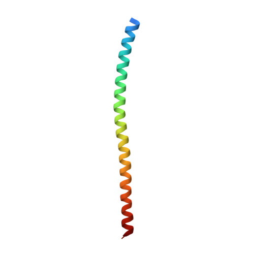
Two members of the B-cell associated 31 (BAP31) family are found in humans; BAP29 and BAP31. These are ubiquitously expressed receptors residing in the endoplasmic reticulum. BAP31 functions in sorting of membrane proteins and in caspase-8 mediated apoptosis, while BAP29 appears to mainly corroborate with BAP31 in sorting. The N-terminal half of these proteins is membrane-bound while the C-terminal half is cytoplasmic. The latter include the so called variant of death effector domain (vDED), which shares weak sequence homology with DED domains. Here we present two structures of BAP31 vDED determined from a single and a twinned crystal, grown at pH 8.0 and pH 4.2, respectively. These structures show that BAP31 vDED forms a dimeric parallel coiled coil with no structural similarity to DED domains. Solution studies support this conclusion and strongly suggest that an additional α-helical domain is present in the C-terminal cytoplasmic region, probably forming a second coiled coil. The thermal stability of BAP31 vDED is quite modest at neutral pH, suggesting that it may assemble in a dynamic fashion in vivo. Surprisingly, BAP29 vDED is partially unfolded at pH 7, while a coiled coil is formed at pH 4.2 in vitro. It is however likely that folding of the domain is triggered by other factors than low pH in vivo. We found no evidence for direct interaction of the cytoplasmic domains of BAP29 and BAP31.
- Department of Medical Biochemistry and Biophysics, Karolinska Institutet, Stockholm, Sweden.
Organizational Affiliation:


