4H1L | pdb_00004h1l

TCR interaction with peptide mimics of nickel offers structural insights in nickel contact allergy


Experimental Data Snapshot

  • Method: X-RAY DIFFRACTION
  • Resolution: 3.30 Å
  • R-Value Free: 
    0.285 (Depositor), 0.261 (DCC) 
  • R-Value Work: 
    0.263 (Depositor), 0.253 (DCC) 
  • R-Value Observed: 
    0.264 (Depositor) 

wwPDB Validation 3D Report Full Report

Validation slider image for 4H1L

This is version 1.2 of the entry. See complete history

Literature

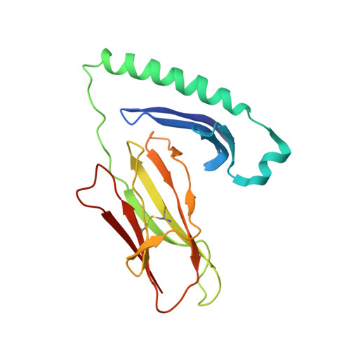
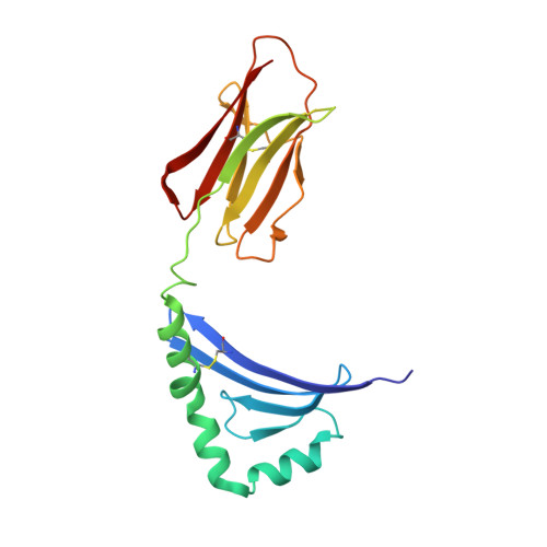
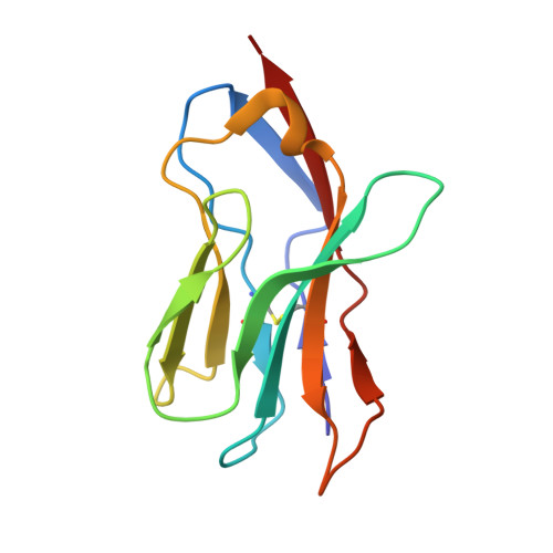
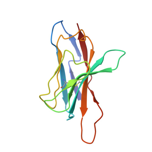
T-cell receptor (TCR) interaction with peptides that mimic nickel offers insight into nickel contact allergy.

Yin, L.Crawford, F.Marrack, P.Kappler, J.W.Dai, S.

(2012) Proc Natl Acad Sci U S A 109: 18517-18522

  • DOI: https://doi.org/10.1073/pnas.1215928109
  • Primary Citation Related Structures: 
    4H1L, 4H25, 4H26

  • PubMed Abstract: 

    T cell-mediated allergy to Ni(++) is one of the most common forms of allergic contact dermatitis, but how the T-cell receptor (TCR) recognizes Ni(++) is unknown. We studied a TCR from an allergic patient that recognizes Ni(++) bound to the MHCII molecule DR52c containing an unknown self-peptide. We identified mimotope peptides that can replace both the self-peptide and Ni(++) in this ligand. They share a p7 lysine whose εNH(2) group is surface-exposed when bound to DR52c. Whereas the TCR uses germ-line complementary-determining region (CDR)1/2 amino acids to dock in the conventional diagonal mode on the mimotope-DR52c complex, the interface is dominated by the TCR Vβ CDR3 interaction with the p7 lysine. Mutations in the TCR CDR loops have similar effects on the T-cell response to either the mimotope or Ni(++) ligand. We suggest that the mimotope p7 lysine mimics Ni(++) in the natural TCR ligand and that MHCII β-chain flexibility in the area around the peptide p7 position forms a common site for cation binding in metal allergies.


  • Organizational Affiliation
    • Howard Hughes Medical Institute, National Jewish Health, Denver, CO 80206, USA.

Macromolecule Content 

  • Total Structure Weight: 138.35 kDa 
  • Atom Count: 9,630 
  • Modeled Residue Count: 1,188 
  • Deposited Residue Count: 1,204 
  • Unique protein chains: 5

Macromolecules

Find similar proteins by:|  3D Structure
Entity ID: 1
MoleculeChains  Sequence LengthOrganismDetailsImage
HLA class II histocompatibility antigen, DR alpha chain
A, D
178Homo sapiensMutation(s): 0 
Gene Names: HLA-DRAHLA-DRA1
UniProt & NIH Common Fund Data Resources
Find proteins for P01903 (Homo sapiens)
Explore P01903 
Go to UniProtKB:  P01903
PHAROS:  P01903
GTEx:  ENSG00000204287 
Entity Groups
Sequence Clusters30% Identity50% Identity70% Identity90% Identity95% Identity100% Identity
UniProt GroupP01903
Sequence Annotations
Expand
Reference Sequence
Find similar proteins by:|  3D Structure
Entity ID: 2
MoleculeChains  Sequence LengthOrganismDetailsImage
MHC class II antigen
B, E
187Homo sapiensMutation(s): 0 
Gene Names: HLA-DRB3
UniProt
Find proteins for P79483 (Homo sapiens)
Explore P79483 
Go to UniProtKB:  P79483
Entity Groups
Sequence Clusters30% Identity50% Identity70% Identity90% Identity95% Identity100% Identity
UniProt GroupP79483
Sequence Annotations
Expand
Reference Sequence
Find similar proteins by:  Sequence   |   3D Structure  
Entity ID: 3
MoleculeChains  Sequence LengthOrganismDetailsImage
mimotope peptide
C, F
13Escherichia coliMutation(s): 0 
Find similar proteins by:|  3D Structure
Entity ID: 4
MoleculeChains  Sequence LengthOrganismDetailsImage
Ani2.3 TCR A chain
G, I
113Escherichia coliMutation(s): 0 
UniProt
Find proteins for L7MTK9 (Escherichia coli)
Explore L7MTK9 
Go to UniProtKB:  L7MTK9
Entity Groups
Sequence Clusters30% Identity50% Identity70% Identity90% Identity95% Identity100% Identity
UniProt GroupL7MTK9
Sequence Annotations
Expand
Reference Sequence
Find similar proteins by:|  3D Structure
Entity ID: 5
MoleculeChains  Sequence LengthOrganismDetailsImage
Ani2.3 TCR B chain
H, J
111Escherichia coliMutation(s): 0 
UniProt
Find proteins for L7MTL0 (Escherichia coli)
Explore L7MTL0 
Go to UniProtKB:  L7MTL0
Entity Groups
Sequence Clusters30% Identity50% Identity70% Identity90% Identity95% Identity100% Identity
UniProt GroupL7MTL0
Sequence Annotations
Expand
Reference Sequence

Experimental Data & Validation

Experimental Data

  • Method: X-RAY DIFFRACTION
  • Resolution: 3.30 Å
  • R-Value Free:  0.285 (Depositor), 0.261 (DCC) 
  • R-Value Work:  0.263 (Depositor), 0.253 (DCC) 
  • R-Value Observed: 0.264 (Depositor) 
Space Group: P 63
Unit Cell:
Length ( Å )Angle ( ˚ )
a = 186.722α = 90
b = 186.722β = 90
c = 166.702γ = 120
Software Package:
Software NamePurpose
HKL-2000data collection
PHASESphasing
PHENIXrefinement
HKL-2000data reduction
HKL-2000data scaling

Structure Validation

View Full Validation Report


Entry History 

Deposition Data

Revision History  (Full details and data files)

  • Version 1.0: 2012-11-14
    Type: Initial release
  • Version 1.1: 2013-09-04
    Changes: Database references
  • Version 1.2: 2024-11-27
    Changes: Data collection, Database references, Structure summary