4GNK | pdb_00004gnk

Crystal structure of Galphaq in complex with full-length human PLCbeta3


Experimental Data Snapshot

  • Method: X-RAY DIFFRACTION
  • Resolution: 4.00 Å
  • R-Value Free: 
    0.255 (Depositor), 0.259 (DCC) 
  • R-Value Work: 
    0.214 (Depositor), 0.222 (DCC) 
  • R-Value Observed: 
    0.216 (Depositor) 

Starting Model: experimental
View more details

wwPDB Validation 3D Report Full Report

Validation slider image for 4GNK

Ligand Structure Quality Assessment 


This is version 1.2 of the entry. See complete history

Literature

Full-length G alpha (q)-phospholipase C-beta 3 structure reveals interfaces of the C-terminal coiled-coil domain.

Lyon, A.M.Dutta, S.Boguth, C.A.Skiniotis, G.Tesmer, J.J.

(2013) Nat Struct Mol Biol 20: 355-362

  • DOI: https://doi.org/10.1038/nsmb.2497
  • Primary Citation Related Structures: 
    4GNK

  • PubMed Abstract: 

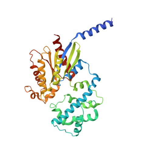
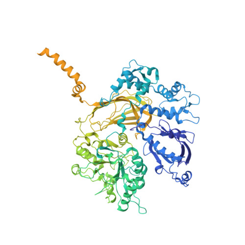
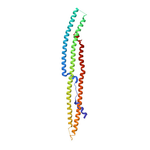
    Phospholipase C-β (PLCβ) is directly activated by Gαq, but the molecular basis for how its distal C-terminal domain (CTD) contributes to maximal activity is poorly understood. Herein we present both the crystal structure and cryo-EM three-dimensional reconstructions of human full-length PLCβ3 in complex with mouse Gαq. The distal CTD forms an extended monomeric helical bundle consisting of three antiparallel segments with structural similarity to membrane-binding bin-amphiphysin-Rvs (BAR) domains. Sequence conservation of the distal CTD suggests putative membrane and protein interaction sites, the latter of which bind the N-terminal helix of Gαq in both the crystal structure and cryo-EM reconstructions. Functional analysis suggests that the distal CTD has roles in membrane targeting and in optimizing the orientation of the catalytic core at the membrane for maximal rates of lipid hydrolysis.


  • Organizational Affiliation
    • Life Sciences Institute, University of Michigan, Ann Arbor, Michigan, USA.

Macromolecule Content 

  • Total Structure Weight: 393.28 kDa 
  • Atom Count: 19,764 
  • Modeled Residue Count: 2,428 
  • Deposited Residue Count: 3,435 
  • Unique protein chains: 3

Macromolecules

Find similar proteins by:|  3D Structure
Entity ID: 1
MoleculeChains  Sequence LengthOrganismDetailsImage
Guanine nucleotide-binding protein G(q) subunit alpha
A, C
353Mus musculusMutation(s): 0 
Gene Names: g alpha qGnaq
EC: 3.6.5
Membrane Entity: Yes 
UniProt
Find proteins for P21279 (Mus musculus)
Explore P21279 
Go to UniProtKB:  P21279
Entity Groups
Sequence Clusters30% Identity50% Identity70% Identity90% Identity95% Identity100% Identity
UniProt GroupP21279
Sequence Annotations
Expand
Reference Sequence
Find similar proteins by:|  3D Structure
Entity ID: 2
MoleculeChains  Sequence LengthOrganismDetailsImage
1-phosphatidylinositol 4,5-bisphosphate phosphodiesterase beta-3
B, D
1,235Homo sapiensMutation(s): 0 
Gene Names: phospholipase c beta 3PLCB3
EC: 3.1.4.11
Membrane Entity: Yes 
UniProt & NIH Common Fund Data Resources
Find proteins for Q01970 (Homo sapiens)
Explore Q01970 
Go to UniProtKB:  Q01970
PHAROS:  Q01970
GTEx:  ENSG00000149782 
Entity Groups
Sequence Clusters30% Identity50% Identity70% Identity90% Identity95% Identity100% Identity
UniProt GroupQ01970
Sequence Annotations
Expand
Reference Sequence
Find similar proteins by:|  3D Structure
Entity ID: 3
MoleculeChains  Sequence LengthOrganismDetailsImage
1-phosphatidylinositol 4,5-bisphosphate phosphodiesterase beta-3259Homo sapiensMutation(s): 0 
Gene Names: phospholipase c beta 3PLCB3
EC: 3.1.4.11
Membrane Entity: Yes 
UniProt & NIH Common Fund Data Resources
Find proteins for Q01970 (Homo sapiens)
Explore Q01970 
Go to UniProtKB:  Q01970
PHAROS:  Q01970
GTEx:  ENSG00000149782 
Entity Groups
Sequence Clusters30% Identity50% Identity70% Identity90% Identity95% Identity100% Identity
UniProt GroupQ01970
Sequence Annotations
Expand
Reference Sequence

Small Molecules

Ligands 4 Unique
IDChains Name / Formula / InChI Key2D Diagram3D Interactions
GDP

Query on GDP


Download:Ideal Coordinates CCD File
F [auth A],
J [auth C]
GUANOSINE-5'-DIPHOSPHATE
C10 H15 N5 O11 P2
QGWNDRXFNXRZMB-UUOKFMHZSA-N
ALF

Query on ALF


Download:Ideal Coordinates CCD File
G [auth A],
K [auth C]
TETRAFLUOROALUMINATE ION
Al F4
UYOMQIYKOOHAMK-UHFFFAOYSA-J
CA

Query on CA


Download:Ideal Coordinates CCD File
I [auth B],
M [auth D]
CALCIUM ION
Ca
BHPQYMZQTOCNFJ-UHFFFAOYSA-N
MG

Query on MG


Download:Ideal Coordinates CCD File
H [auth A],
L [auth C]
MAGNESIUM ION
Mg
JLVVSXFLKOJNIY-UHFFFAOYSA-N

Experimental Data & Validation

Experimental Data

  • Method: X-RAY DIFFRACTION
  • Resolution: 4.00 Å
  • R-Value Free:  0.255 (Depositor), 0.259 (DCC) 
  • R-Value Work:  0.214 (Depositor), 0.222 (DCC) 
  • R-Value Observed: 0.216 (Depositor) 
Space Group: P 21 21 21
Unit Cell:
Length ( Å )Angle ( ˚ )
a = 91.423α = 90
b = 188.845β = 90
c = 293.857γ = 90
Software Package:
Software NamePurpose
HKL-2000data collection
PHASERphasing
REFMACrefinement
DENZOdata reduction
SCALEPACKdata scaling

Structure Validation

View Full Validation Report


Ligand Structure Quality Assessment 


Entry History 

Deposition Data

Revision History  (Full details and data files)

  • Version 1.0: 2013-02-06
    Type: Initial release
  • Version 1.1: 2013-09-04
    Changes: Database references
  • Version 1.2: 2023-09-13
    Changes: Data collection, Database references, Derived calculations, Refinement description