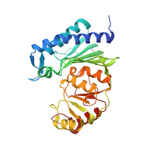
4G61

Crystal structure of IMPase/NADP phosphatase complexed with Mg2+ and phosphate


Experimental Data Snapshot

  • Method: X-RAY DIFFRACTION
  • Resolution: 2.30 Å
  • R-Value Free: 0.255 
  • R-Value Work: 0.176 
  • R-Value Observed: 0.180 

wwPDB Validation   3D Report Full Report


Ligand Structure Quality Assessment 


This is version 1.2 of the entry. See complete history


Literature

Structural elucidation of the binding site and mode of inhibition of Li(+) and Mg(2+) in inositol monophosphatase.

Dutta, A.Bhattacharyya, S.Dutta, D.Das, A.K.

(2014) FEBS J 281: 5309-5324

  • DOI: https://doi.org/10.1111/febs.13070
  • Primary Citation of Related Structures:  
    4G61, 4I3Y, 4I40, 4PTK

  • PubMed Abstract: 

    Mg(2+) -dependent, Li(+) -sensitive phosphatases are a widely distributed family of enzymes with significant importance throughout the biological kingdom. Inositol monophosphatase (IMPase) is an important target of Li(+) -based therapeutic agents in manic depressive disorders. However, despite decades of intense research efforts, the precise mechanism of Li(+) -induced inhibition of IMPase remains obscured. Here we describe a structural investigation of the Li(+) binding site in staphylococcal IMPase I (SaIMPase I) using X-ray crystallography. The biochemical study indicated common or overlapping binding sites for Mg(2+) and Li(+) in the active site of SaIMPase I. The crystal structure of the SaIMPase I ternary product complex shows the presence of a phosphate and three Mg(2+) ions (namely Mg1, Mg2 and Mg3) in the active site. As Li(+) is virtually invisible in X-ray crystallography, competitive displacement of Mg(2+) ions from the SaIMPase I ternary product complex as a function of increasing LiCl concentration was used to identify the Li(+) binding site. In this approach, the disappearing electron density of Mg(2+) ions due to Li(+) ion binding was traced, and the Mg(2+) ion present at the Mg2 binding site was found to be replaced. Moreover, based on a detailed comparative investigation of the phosphate orientation and coordination states of Mg(2+) binding sites in enzyme-substrate and enzyme-product complexes, inhibition mechanisms for Li(+) and Mg(2+) are proposed.


  • Organizational Affiliation

    Department of Biotechnology, Indian Institute of Technology Kharagpur, Kharagpur, West Bengal, India.


Macromolecules
Find similar proteins by:  (by identity cutoff)  |  3D Structure
Entity ID: 1
MoleculeChains Sequence LengthOrganismDetailsImage
Inositol monophosphatase family protein
A, B
271Staphylococcus aureus subsp. aureus MSSA476Mutation(s): 0 
Gene Names: SAS2203
EC: 3.1.3.25
Entity Groups  
Sequence Clusters30% Identity50% Identity70% Identity90% Identity95% Identity100% Identity
Sequence Annotations
Expand
  • Reference Sequence
Small Molecules
Ligands 7 Unique
IDChains Name / Formula / InChI Key2D Diagram3D Interactions
P33
Query on P33

Download Ideal Coordinates CCD File 
R [auth B]3,6,9,12,15,18-HEXAOXAICOSANE-1,20-DIOL
C14 H30 O8
XPJRQAIZZQMSCM-UHFFFAOYSA-N
PG4
Query on PG4

Download Ideal Coordinates CCD File 
S [auth B]TETRAETHYLENE GLYCOL
C8 H18 O5
UWHCKJMYHZGTIT-UHFFFAOYSA-N
DTO
Query on DTO

Download Ideal Coordinates CCD File 
P [auth B]1-HYDROXYSULFANYL-4-MERCAPTO-BUTANE-2,3-DIOL
C4 H10 O3 S2
MFLGZMZEMVZQCC-QWWZWVQMSA-N
PO4
Query on PO4

Download Ideal Coordinates CCD File 
E [auth A],
J [auth B]
PHOSPHATE ION
O4 P
NBIIXXVUZAFLBC-UHFFFAOYSA-K
GOL
Query on GOL

Download Ideal Coordinates CCD File 
G [auth A],
I [auth A],
N [auth B],
Q [auth B]
GLYCEROL
C3 H8 O3
PEDCQBHIVMGVHV-UHFFFAOYSA-N
CL
Query on CL

Download Ideal Coordinates CCD File 
H [auth A],
O [auth B]
CHLORIDE ION
Cl
VEXZGXHMUGYJMC-UHFFFAOYSA-M
MG
Query on MG

Download Ideal Coordinates CCD File 
C [auth A]
D [auth A]
F [auth A]
K [auth B]
L [auth B]
C [auth A],
D [auth A],
F [auth A],
K [auth B],
L [auth B],
M [auth B]
MAGNESIUM ION
Mg
JLVVSXFLKOJNIY-UHFFFAOYSA-N
Experimental Data & Validation

Experimental Data

  • Method: X-RAY DIFFRACTION
  • Resolution: 2.30 Å
  • R-Value Free: 0.255 
  • R-Value Work: 0.176 
  • R-Value Observed: 0.180 
  • Space Group: P 21 2 21
Unit Cell:
Length ( Å )Angle ( ˚ )
a = 60.709α = 90
b = 63.054β = 90
c = 141.617γ = 90
Software Package:
Software NamePurpose
SCALAdata scaling
MOLREPphasing
REFMACrefinement
PDB_EXTRACTdata extraction
StructureStudiodata collection
XSCALEdata scaling

Structure Validation

View Full Validation Report


Ligand Structure Quality Assessment 


Entry History 

Deposition Data

Revision History  (Full details and data files)

  • Version 1.0: 2013-07-24
    Type: Initial release
  • Version 1.1: 2014-10-22
    Changes: Database references
  • Version 1.2: 2022-08-24
    Changes: Database references, Derived calculations, Structure summary