Structural elucidation of the binding site and mode of inhibition of Li(+) and Mg(2+) in inositol monophosphatase.
Dutta, A., Bhattacharyya, S., Dutta, D., Das, A.K.(2014) FEBS J 281: 5309-5324
- PubMed: 25263816 Search on PubMed
- DOI: https://doi.org/10.1111/febs.13070
- Primary Citation Related Structures:
4G61, 4I3Y, 4I40, 4PTK - PubMed Abstract:
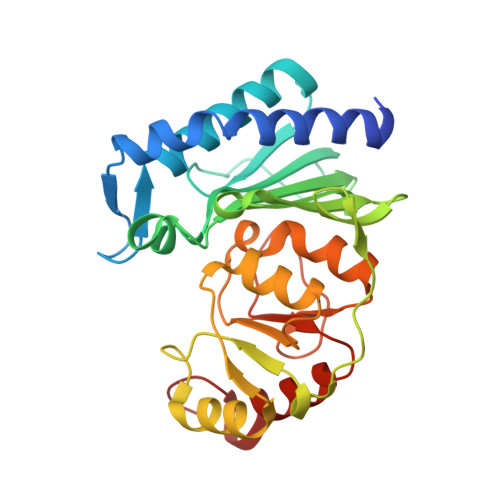
Mg(2+) -dependent, Li(+) -sensitive phosphatases are a widely distributed family of enzymes with significant importance throughout the biological kingdom. Inositol monophosphatase (IMPase) is an important target of Li(+) -based therapeutic agents in manic depressive disorders. However, despite decades of intense research efforts, the precise mechanism of Li(+) -induced inhibition of IMPase remains obscured. Here we describe a structural investigation of the Li(+) binding site in staphylococcal IMPase I (SaIMPase I) using X-ray crystallography. The biochemical study indicated common or overlapping binding sites for Mg(2+) and Li(+) in the active site of SaIMPase I. The crystal structure of the SaIMPase I ternary product complex shows the presence of a phosphate and three Mg(2+) ions (namely Mg1, Mg2 and Mg3) in the active site. As Li(+) is virtually invisible in X-ray crystallography, competitive displacement of Mg(2+) ions from the SaIMPase I ternary product complex as a function of increasing LiCl concentration was used to identify the Li(+) binding site. In this approach, the disappearing electron density of Mg(2+) ions due to Li(+) ion binding was traced, and the Mg(2+) ion present at the Mg2 binding site was found to be replaced. Moreover, based on a detailed comparative investigation of the phosphate orientation and coordination states of Mg(2+) binding sites in enzyme-substrate and enzyme-product complexes, inhibition mechanisms for Li(+) and Mg(2+) are proposed. The atomic coordinates for the SaIMPase I ternary complex, SaIMPase I in 50 mm LiCl, SaIMPase I in 100 mm LiCl and SaIMPase I in 0 mm MgCl2 have been submitted to the Protein Data Bank under accession numbers 4G61, 4I40, 4I3Y and 4PTK, respectively.
- Department of Biotechnology, Indian Institute of Technology Kharagpur, Kharagpur, West Bengal, India.
Organizational Affiliation: