


DETERMINATION of SITE AFFINITY CONSTANTS FOR PRODUCT INHIBITION OF WILD-TYPE AND MUTANT FORMS OF RECOMBINANT HUMAN HEXOKINASE TYPE I
Shen, L., Gao, Y., Honzatko, R.B.To be published.
Experimental Data Snapshot
Entity ID: 1 | |||||
|---|---|---|---|---|---|
| Molecule | Chains | Sequence Length | Organism | Details | Image |
| Hexokinase-1 | 917 | Homo sapiens | Mutation(s): 1 Gene Names: HK1 EC: 2.7.1.1 | 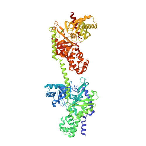 | |
UniProt & NIH Common Fund Data Resources | |||||
PHAROS: P19367 GTEx: ENSG00000156515 | |||||
Entity Groups | |||||
| Sequence Clusters | 30% Identity50% Identity70% Identity90% Identity95% Identity100% Identity | ||||
| UniProt Group | P19367 | ||||
Sequence AnnotationsExpand | |||||
Reference Sequence | |||||
| Ligands 4 Unique | |||||
|---|---|---|---|---|---|
| ID | Chains | Name / Formula / InChI Key | 2D Diagram | 3D Interactions | |
| G16 Download:Ideal Coordinates CCD File | D [auth A], F [auth A], K [auth B], M [auth B] | 1,6-di-O-phosphono-alpha-D-glucopyranose C6 H13 O12 P2 RWHOZGRAXYWRNX-VFUOTHLCSA-M |  | ||
| CIT Download:Ideal Coordinates CCD File | I [auth A], P [auth B] | CITRIC ACID C6 H8 O7 KRKNYBCHXYNGOX-UHFFFAOYSA-N |  | ||
| BGC Download:Ideal Coordinates CCD File | C [auth A], E [auth A], J [auth B], L [auth B] | beta-D-glucopyranose C6 H12 O6 WQZGKKKJIJFFOK-VFUOTHLCSA-N |  | ||
| NA Download:Ideal Coordinates CCD File | G [auth A], H [auth A], N [auth B], O [auth B] | SODIUM ION Na FKNQFGJONOIPTF-UHFFFAOYSA-N |  | ||
| Length ( Å ) | Angle ( ˚ ) |
|---|---|
| a = 82.38 | α = 90 |
| b = 120.772 | β = 92.8 |
| c = 120.591 | γ = 90 |
| Software Name | Purpose |
|---|---|
| REFMAC | refinement |
| PDB_EXTRACT | data extraction |
| HKL-3000 | data collection |
| HKL-3000 | data reduction |
| HKL-3000 | data scaling |
| AMoRE | phasing |


