Structure and mechanism of a bacterial sodium-dependent dicarboxylate transporter.
Mancusso, R., Gregorio, G.G., Liu, Q., Wang, D.N.(2012) Nature 491: 622-626
- PubMed: 23086149 Search on PubMedSearch on PubMed Central
- DOI: https://doi.org/10.1038/nature11542
- Primary Citation Related Structures:
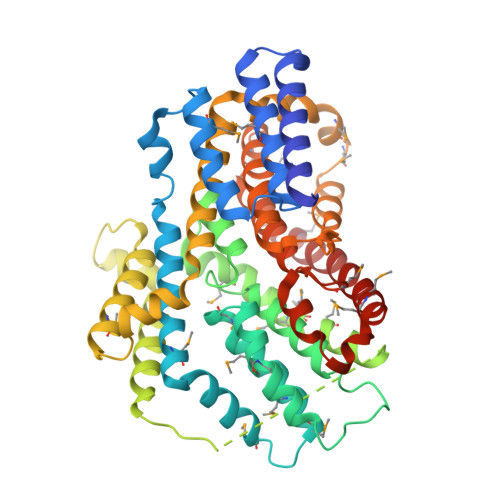
4F35 - PubMed Abstract:
In human cells, cytosolic citrate is a chief precursor for the synthesis of fatty acids, triacylglycerols, cholesterol and low-density lipoprotein. Cytosolic citrate further regulates the energy balance of the cell by activating the fatty-acid-synthesis pathway while downregulating both the glycolysis and fatty-acid β-oxidation pathways. The rate of fatty-acid synthesis in liver and adipose cells, the two main tissue types for such synthesis, correlates directly with the concentration of citrate in the cytosol, with the cytosolic citrate concentration partially depending on direct import across the plasma membrane through the Na(+)-dependent citrate transporter (NaCT). Mutations of the homologous fly gene (Indy; I'm not dead yet) result in reduced fat storage through calorie restriction. More recently, Nact (also known as Slc13a5)-knockout mice have been found to have increased hepatic mitochondrial biogenesis, higher lipid oxidation and energy expenditure, and reduced lipogenesis, which taken together protect the mice from obesity and insulin resistance. To understand the transport mechanism of NaCT and INDY proteins, here we report the 3.2 Å crystal structure of a bacterial INDY homologue. One citrate molecule and one sodium ion are bound per protein, and their binding sites are defined by conserved amino acid motifs, forming the structural basis for understanding the specificity of the transporter. Comparison of the structures of the two symmetrical halves of the transporter suggests conformational changes that propel substrate translocation.
- The Helen L. and Martin S. Kimmel Center for Biology and Medicine at the Skirball Institute of Biomolecular Medicine, New York University School of Medicine, 540 First Avenue, New York, New York 10016, USA.
Organizational Affiliation: