4EMQ | pdb_00004emq

Crystal structure of a single mutant of Dronpa, the green-on-state PDM1-4


Experimental Data Snapshot

  • Method: X-RAY DIFFRACTION
  • Resolution: 1.95 Å
  • R-Value Free: 
    0.232 (Depositor), 0.227 (DCC) 
  • R-Value Work: 
    0.183 (Depositor), 0.180 (DCC) 
  • R-Value Observed: 
    0.186 (Depositor) 

Starting Model: experimental
View more details

wwPDB Validation 3D Report Full Report

Validation slider image for 4EMQ

This is version 2.1 of the entry. See complete history

Literature

Structural basis for the influence of a single mutation K145N on the oligomerization and photoswitching rate of Dronpa.

Nguyen Bich, N.Moeyaert, B.Van Hecke, K.Dedecker, P.Mizuno, H.Hofkens, J.Van Meervelt, L.

(2012) Acta Crystallogr D Biol Crystallogr 68: 1653-1659

  • DOI: https://doi.org/10.1107/S0907444912039686
  • Primary Citation Related Structures: 
    4EMQ

  • PubMed Abstract: 

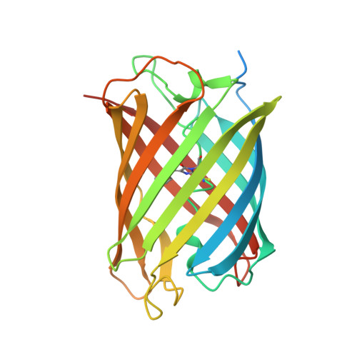
    The crystal structure of the on-state of PDM1-4, a single-mutation variant of the photochromic fluorescent protein Dronpa, is reported at 1.95 Å resolution. PDM1-4 is a Dronpa variant that possesses a slower off-switching rate than Dronpa and thus can effectively increase the image resolution in subdiffraction optical microscopy, although the precise molecular basis for this change has not been elucidated. This work shows that the Lys145Asn mutation in PDM1-4 stabilizes the interface available for dimerization, facilitating oligomerization of the protein. No significant changes were observed in the chromophore environment of PDM1-4 compared with Dronpa, and the ensemble absorption and emission properties of PDM1-4 were highly similar to those of Dronpa. It is proposed that the slower off-switching rate in PDM1-4 is caused by a decrease in the potential flexibility of certain β-strands caused by oligomerization along the AC interface.


  • Organizational Affiliation
    • Biomolecular Architecture, Department of Chemistry, KU Leuven, Celestijnenlaan 200F, Leuven (Heverlee), Belgium.

Macromolecule Content 

  • Total Structure Weight: 177.8 kDa 
  • Atom Count: 11,412 
  • Modeled Residue Count: 1,286 
  • Deposited Residue Count: 1,530 
  • Unique protein chains: 1

Macromolecules

Find similar proteins by:|  3D Structure
Entity ID: 1
MoleculeChains  Sequence LengthOrganismDetailsImage
Fluorescent protein Dronpa
A, B, C, D, E
A, B, C, D, E, F
255Echinophyllia sp. SC22Mutation(s): 2 
Gene Names: Dronpa
UniProt
Find proteins for Q5TLG6 (Echinophyllia sp. SC22)
Explore Q5TLG6 
Go to UniProtKB:  Q5TLG6
Entity Groups
Sequence Clusters30% Identity50% Identity70% Identity90% Identity95% Identity100% Identity
UniProt GroupQ5TLG6
Sequence Annotations
Expand
Reference Sequence

Small Molecules

Ligands 3 Unique
IDChains Name / Formula / InChI Key2D Diagram3D Interactions
EPE

Query on EPE


Download:Ideal Coordinates CCD File
G [auth A]
I [auth B]
K [auth C]
N [auth D]
P [auth E]
G [auth A],
I [auth B],
K [auth C],
N [auth D],
P [auth E],
R [auth F]
4-(2-HYDROXYETHYL)-1-PIPERAZINE ETHANESULFONIC ACID
C8 H18 N2 O4 S
JKMHFZQWWAIEOD-UHFFFAOYSA-N
1PE

Query on 1PE


Download:Ideal Coordinates CCD File
H [auth A]PENTAETHYLENE GLYCOL
C10 H22 O6
JLFNLZLINWHATN-UHFFFAOYSA-N
PEG

Query on PEG


Download:Ideal Coordinates CCD File
J [auth B]
L [auth C]
M [auth C]
O [auth D]
Q [auth E]
J [auth B],
L [auth C],
M [auth C],
O [auth D],
Q [auth E],
S [auth F],
T [auth F]
DI(HYDROXYETHYL)ETHER
C4 H10 O3
MTHSVFCYNBDYFN-UHFFFAOYSA-N
Modified Residues  1 Unique
IDChains TypeFormula2D DiagramParent
GYC
Query on GYC
A, B, C, D, E
A, B, C, D, E, F
L-PEPTIDE LINKINGC14 H15 N3 O4 SCYS, TYR, GLY

Experimental Data & Validation

Experimental Data

  • Method: X-RAY DIFFRACTION
  • Resolution: 1.95 Å
  • R-Value Free:  0.232 (Depositor), 0.227 (DCC) 
  • R-Value Work:  0.183 (Depositor), 0.180 (DCC) 
  • R-Value Observed: 0.186 (Depositor) 
Space Group: P 2 21 21
Unit Cell:
Length ( Å )Angle ( ˚ )
a = 69.539α = 90
b = 103.804β = 90
c = 177.255γ = 90
Software Package:
Software NamePurpose
XDSdata scaling
PHASESphasing
PHENIXrefinement
XDSdata reduction
SCALAdata scaling

Structure Validation

View Full Validation Report


Entry History 

Deposition Data

Revision History  (Full details and data files)

  • Version 1.0: 2012-11-21
    Type: Initial release
  • Version 1.1: 2013-01-23
    Changes: Database references
  • Version 1.2: 2023-09-13
    Changes: Data collection, Database references, Derived calculations, Refinement description
  • Version 2.0: 2023-11-15
    Changes: Advisory, Atomic model, Data collection, Derived calculations
  • Version 2.1: 2024-10-09
    Changes: Structure summary