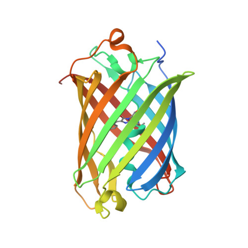
Structural basis for bathochromic shift of fluorescence in far-red fluorescent proteins eqFP650 and eqFP670.
Pletnev, S., Pletneva, N.V., Souslova, E.A., Chudakov, D.M., Lukyanov, S., Wlodawer, A., Dauter, Z., Pletnev, V.(2012) Acta Crystallogr D Biol Crystallogr 68: 1088-1097
- PubMed: 22948909 Search on PubMedSearch on PubMed Central
- DOI: https://doi.org/10.1107/S0907444912020598
- Primary Citation Related Structures:
4EDO, 4EDS - PubMed Abstract:
The crystal structures of the far-red fluorescent proteins (FPs) eqFP650 (λ(ex)(max)/λ(em)(max) 592/650 nm) and eqFP670 (λ(ex)(max)/λ(em)(max) 605/670 nm), the successors of the far-red FP Katushka (λ(ex)(max)/λ(em)(max) 588/635 nm), have been determined at 1.8 and 1.6 Å resolution, respectively. An examination of the structures demonstrated that there are two groups of changes responsible for the bathochromic shift of excitation/emission bands of these proteins relative to their predecessor. The first group of changes resulted in an increase of hydrophilicity at the acylimine site of the chromophore due to the presence of one and three water molecules in eqFP650 and eqFP670, respectively. These water molecules provide connection of the chromophore with the protein scaffold via hydrogen bonds causing an ~15 nm bathochromic shift of the eqFP650 and eqFP670 emission bands. The second group of changes observed in eqFP670 arises from substitution of both Ser143 and Ser158 by asparagines. Asn143 and Asn158 of eqFP670 are hydrogen bonded with each other, as well as with the protein scaffold and with the p-hydroxyphenyl group of the chromophore, resulting in an additional ~20 nm bathochromic shift of the eqFP670 emission band as compared to eqFP650. The role of the observed structural changes was verified by mutagenesis.
- Synchrotron Radiation Research Section, Macromolecular Crystallography Laboratory, National Cancer Institute, Argonne, IL 60439, USA. pletnevs@mail.nih.gov
Organizational Affiliation: