4E06 | pdb_00004e06

Anophelin from the malaria vector inhibits thrombin through a novel reverse-binding mechanism


Experimental Data Snapshot

  • Method: X-RAY DIFFRACTION
  • Resolution: 3.20 Å
  • R-Value Free: 
    0.208 (Depositor), 0.205 (DCC) 
  • R-Value Work: 
    0.177 (Depositor), 0.178 (DCC) 
  • R-Value Observed: 
    0.178 (Depositor) 

Starting Model: experimental
View more details

wwPDB Validation 3D Report Full Report

Validation slider image for 4E06

Ligand Structure Quality Assessment 


This is version 1.5 of the entry. See complete history

Literature

Unique thrombin inhibition mechanism by anophelin, an anticoagulant from the malaria vector.

Figueiredo, A.C.de Sanctis, D.Gutierrez-Gallego, R.Cereija, T.B.Macedo-Ribeiro, S.Fuentes-Prior, P.Pereira, P.J.

(2012) Proc Natl Acad Sci U S A 109: E3649-E3658

  • DOI: https://doi.org/10.1073/pnas.1211614109
  • Primary Citation Related Structures: 
    4E05, 4E06

  • PubMed Abstract: 

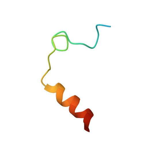
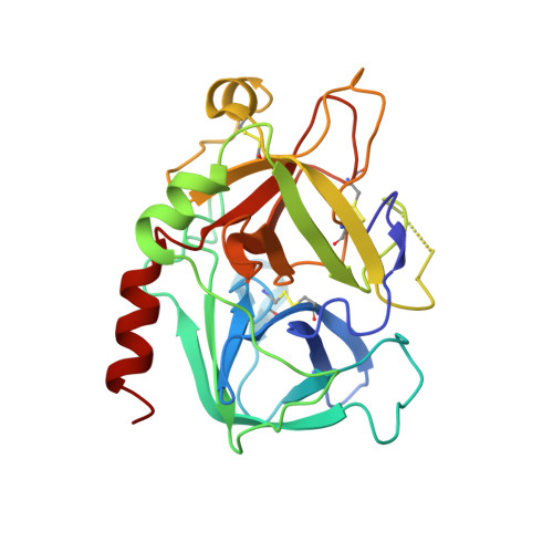
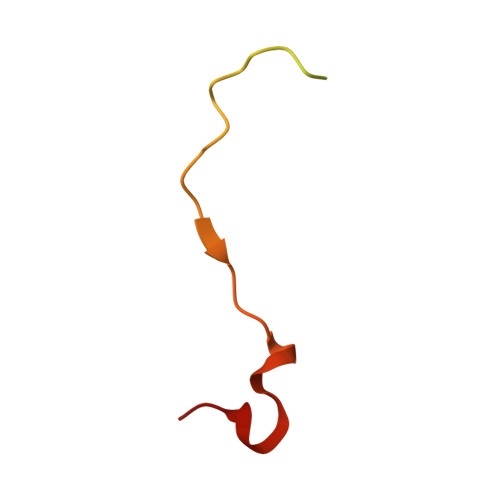
    Anopheles mosquitoes are vectors of malaria, a potentially fatal blood disease affecting half a billion humans worldwide. These blood-feeding insects include in their antihemostatic arsenal a potent thrombin inhibitor, the flexible and cysteine-less anophelin. Here, we present a thorough structure-and-function analysis of thrombin inhibition by anophelin, including the 2.3-Å crystal structure of the human thrombin·anophelin complex. Anophelin residues 32-61 are well-defined by electron density, completely occupying the long cleft between the active site and exosite I. However, in striking contrast to substrates, the D50-R53 anophelin tetrapeptide occupies the active site cleft of the enzyme, whereas the upstream residues A35-P45 shield the regulatory exosite I, defining a unique reverse-binding mode of an inhibitor to the target proteinase. The extensive interactions established, the disruption of thrombin's active site charge-relay system, and the insertion of residue R53 into the proteinase S(1) pocket in an orientation opposed to productive substrates explain anophelin's remarkable specificity and resistance to proteolysis by thrombin. Complementary biophysical and functional characterization of point mutants and truncated versions of anophelin unambiguously establish the molecular mechanism of action of this family of serine proteinase inhibitors (I77). These findings have implications for the design of novel antithrombotics.


  • Organizational Affiliation
    • Instituto de Biologia Molecular e Celular (IBMC), Universidade do Porto, 4150-180 Porto, Portugal.

Macromolecule Content 

  • Total Structure Weight: 40.66 kDa 
  • Atom Count: 2,512 
  • Modeled Residue Count: 308 
  • Deposited Residue Count: 356 
  • Unique protein chains: 3

Macromolecules

Find similar proteins by:|  3D Structure
Entity ID: 1
MoleculeChains  Sequence LengthOrganismDetailsImage
ThrombinA [auth L]36Homo sapiensMutation(s): 0 
EC: 3.4.21.5
UniProt & NIH Common Fund Data Resources
Find proteins for P00734 (Homo sapiens)
Explore P00734 
Go to UniProtKB:  P00734
PHAROS:  P00734
GTEx:  ENSG00000180210 
Entity Groups
Sequence Clusters30% Identity50% Identity70% Identity90% Identity95% Identity100% Identity
UniProt GroupP00734
Sequence Annotations
Expand
Reference Sequence
Find similar proteins by:|  3D Structure
Entity ID: 2
MoleculeChains  Sequence LengthOrganismDetailsImage
ThrombinB [auth H]259Homo sapiensMutation(s): 0 
EC: 3.4.21.5
UniProt & NIH Common Fund Data Resources
Find proteins for P00734 (Homo sapiens)
Explore P00734 
Go to UniProtKB:  P00734
PHAROS:  P00734
GTEx:  ENSG00000180210 
Entity Groups
Sequence Clusters30% Identity50% Identity70% Identity90% Identity95% Identity100% Identity
UniProt GroupP00734
Glycosylation
Glycosylation Sites: 1Go to GlyGen: P00734-1
Sequence Annotations
Expand
Reference Sequence
Find similar proteins by:|  3D Structure
Entity ID: 3
MoleculeChains  Sequence LengthOrganismDetailsImage
Salivary anti-thrombin peptide anophelinC [auth I]61Anopheles albimanusMutation(s): 0 
Gene Names: anophelin
UniProt
Find proteins for Q9NJS1 (Anopheles albimanus)
Explore Q9NJS1 
Go to UniProtKB:  Q9NJS1
Entity Groups
Sequence Clusters30% Identity50% Identity70% Identity90% Identity95% Identity100% Identity
UniProt GroupQ9NJS1
Sequence Annotations
Expand
Reference Sequence

Small Molecules

Experimental Data & Validation

Experimental Data

  • Method: X-RAY DIFFRACTION
  • Resolution: 3.20 Å
  • R-Value Free:  0.208 (Depositor), 0.205 (DCC) 
  • R-Value Work:  0.177 (Depositor), 0.178 (DCC) 
  • R-Value Observed: 0.178 (Depositor) 
Space Group: H 3 2
Unit Cell:
Length ( Å )Angle ( ˚ )
a = 209.659α = 90
b = 209.659β = 90
c = 127.412γ = 120
Software Package:
Software NamePurpose
SCALAdata scaling
PHASERphasing
PHENIXrefinement
PDB_EXTRACTdata extraction
DNAdata collection
XDSdata reduction

Structure Validation

View Full Validation Report


Ligand Structure Quality Assessment 


Entry History 

Deposition Data

Revision History  (Full details and data files)

  • Version 1.0: 2012-12-05
    Type: Initial release
  • Version 1.1: 2012-12-26
    Changes: Database references
  • Version 1.2: 2013-01-09
    Changes: Database references
  • Version 1.3: 2020-07-29
    Type: Remediation
    Reason: Carbohydrate remediation
    Changes: Data collection, Derived calculations, Structure summary
  • Version 1.4: 2023-09-13
    Changes: Data collection, Database references, Refinement description, Structure summary
  • Version 1.5: 2024-10-16
    Changes: Structure summary