4BWC | pdb_00004bwc

X-ray structure of a phospholiapse B like protein 1 from bovine kidneys


Experimental Data Snapshot

  • Method: X-RAY DIFFRACTION
  • Resolution: 1.89 Å
  • R-Value Free: 
    0.231 (Depositor), 0.229 (DCC) 
  • R-Value Work: 
    0.188 (Depositor), 0.187 (DCC) 
  • R-Value Observed: 
    0.190 (Depositor) 

Starting Model: experimental
View more details

wwPDB Validation 3D Report Full Report

Validation slider image for 4BWC

Ligand Structure Quality Assessment 


This is version 2.1 of the entry. See complete history

Literature

Is the Bovine Lysosomal Phospholipase B-Like Protein an Amidase?

Repo, H.Kuokkanen, E.Oksanen, E.Goldman, A.Heikinheimo, P.

(2014) Proteins 82: 300

  • DOI: https://doi.org/10.1002/prot.24388
  • Primary Citation Related Structures: 
    4BWC

  • PubMed Abstract: 

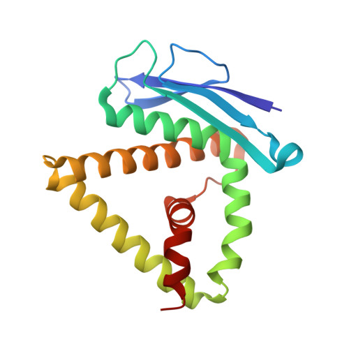
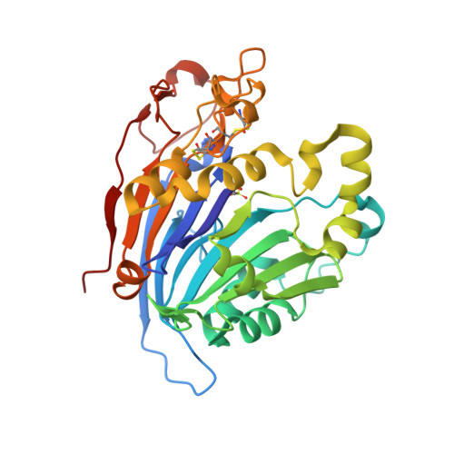
    The main function of lysosomal proteins is to degrade cellular macromolecules. We purified a novel lysosomal protein to homogeneity from bovine kidneys. By gene annotation, this protein is defined as a bovine phospholipase B-like protein 1 (bPLBD1) and, to better understand its biological function, we solved its structure at 1.9 Å resolution. We showed that bPLBD1 has uniform noncomplex-type N-glycosylation and that it localized to the lysosome. The first step in lysosomal protein transport, the initiation of mannose-6-phosphorylation by a N-acetylglucosamine-1-phosphotransferase, requires recognition of at least two distinct lysines on the protein surface. We identified candidate lysines by analyzing the structural and sequentially conserved N-glycosylation sites and lysines in bPLBD1 and in the homologous mouse PLBD2. Our model suggests that N408 is the primarily phosphorylated glycan, and K358 a key residue for N-acetylglucosamine-1-phosphotransferase recognition. Two other lysines, K334 and K342, provide the required second site for N-acetylglucosamine-1-phosphotransferase recognition. bPLBD1 is an N-terminal nucleophile (Ntn) hydrolase. By comparison with other Ntn-hydrolases, we conclude that the acyl moiety of PLBD1 substrate must be small to fit the putative binding pocket, whereas the space for the rest of the substrate is a large open cleft. Finally, as all the known substrates of Ntn-hydrolases have amide bonds, we suggest that bPLBD1 may be an amidase or peptidase instead of lipase, explaining the difficulty in finding a good substrate for any members of the PLBD family.


  • Organizational Affiliation
    • Institute of Biotechnology, University of Helsinki, FI-00014, Helsinki, Finland; Department of Biosciences, University of Helsinki, FI-00014, Helsinki, Finland.

Macromolecule Content 

  • Total Structure Weight: 58.88 kDa 
  • Atom Count: 4,334 
  • Modeled Residue Count: 491 
  • Deposited Residue Count: 491 
  • Unique protein chains: 2

Macromolecules

Find similar proteins by:|  3D Structure
Entity ID: 1
MoleculeChains  Sequence LengthOrganismDetailsImage
PHOSPHOLIPASE B-LIKE 1170Bos taurusMutation(s): 0 
EC: 3.1.1 (PDB Primary Data), 3.4.11 (UniProt)
UniProt
Find proteins for Q9GL30 (Bos taurus)
Explore Q9GL30 
Go to UniProtKB:  Q9GL30
Entity Groups
Sequence Clusters30% Identity50% Identity70% Identity90% Identity95% Identity100% Identity
UniProt GroupQ9GL30
Glycosylation
Glycosylation Sites: 1
Sequence Annotations
Expand
Reference Sequence
Find similar proteins by:|  3D Structure
Entity ID: 2
MoleculeChains  Sequence LengthOrganismDetailsImage
PHOSPHOLIPASE B-LIKE 1321Bos taurusMutation(s): 0 
EC: 3.1.1 (PDB Primary Data), 3.4.11 (UniProt)
UniProt
Find proteins for Q9GL30 (Bos taurus)
Explore Q9GL30 
Go to UniProtKB:  Q9GL30
Entity Groups
Sequence Clusters30% Identity50% Identity70% Identity90% Identity95% Identity100% Identity
UniProt GroupQ9GL30
Glycosylation
Glycosylation Sites: 4
Sequence Annotations
Expand
Reference Sequence

Oligosaccharides

Help  
Entity ID: 3
MoleculeChains Length2D Diagram GlycosylationD Interactions
2-acetamido-2-deoxy-beta-D-glucopyranose-(1-4)-2-acetamido-2-deoxy-beta-D-glucopyranose
C, D, E
2N-Glycosylation
Glycosylation Resources
GlyTouCan: G42666HT
GlyCosmos: G42666HT
GlyGen: G42666HT

Small Molecules

Modified Residues  1 Unique
IDChains TypeFormula2D DiagramParent
OCS
Query on OCS
B
L-PEPTIDE LINKINGC3 H7 N O5 SCYS

Experimental Data & Validation

Experimental Data

  • Method: X-RAY DIFFRACTION
  • Resolution: 1.89 Å
  • R-Value Free:  0.231 (Depositor), 0.229 (DCC) 
  • R-Value Work:  0.188 (Depositor), 0.187 (DCC) 
  • R-Value Observed: 0.190 (Depositor) 
Space Group: P 31 2 1
Unit Cell:
Length ( Å )Angle ( ˚ )
a = 97.37α = 90
b = 97.37β = 90
c = 140.945γ = 120
Software Package:
Software NamePurpose
REFMACrefinement
XDSdata reduction
MOLREPphasing

Structure Validation

View Full Validation Report


Ligand Structure Quality Assessment 


Entry History 

Deposition Data

Revision History  (Full details and data files)

  • Version 1.0: 2013-09-18
    Type: Initial release
  • Version 1.1: 2014-02-05
    Changes: Database references
  • Version 2.0: 2020-07-29
    Type: Remediation
    Reason: Carbohydrate remediation
    Changes: Atomic model, Data collection, Derived calculations, Other, Structure summary
  • Version 2.1: 2023-12-20
    Changes: Data collection, Database references, Refinement description, Structure summary