4AEJ

Crystal structure of Human fibrillar procollagen type III C- propeptide trimer


Experimental Data Snapshot

  • Method: X-RAY DIFFRACTION
  • Resolution: 2.21 Å
  • R-Value Free: 0.237 
  • R-Value Work: 0.201 
  • R-Value Observed: 0.203 

wwPDB Validation   3D Report Full Report


This is version 1.2 of the entry. See complete history


Literature

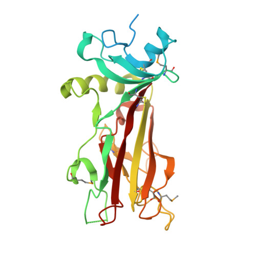
Structural Basis of Fibrillar Collagen Trimerization and Related Genetic Disorders.

Bourhis, J.M.Mariano, N.Zhao, Y.Harlos, K.Exposito, J.Jones, E.Y.Moali, C.Aghajari, N.Hulmes, D.J.S.

(2012) Nat Struct Mol Biol 19: 1031

  • DOI: https://doi.org/10.1038/nsmb.2389
  • Primary Citation of Related Structures:  
    4AE2, 4AEJ, 4AK3

  • PubMed Abstract: 

    The C propeptides of fibrillar procollagens have crucial roles in tissue growth and repair by controlling both the intracellular assembly of procollagen molecules and the extracellular assembly of collagen fibrils. Mutations in C propeptides are associated with several, often lethal, genetic disorders affecting bone, cartilage, blood vessels and skin. Here we report the crystal structure of a C-propeptide domain from human procollagen III. It reveals an exquisite structural mechanism of chain recognition during intracellular trimerization of the procollagen molecule. It also gives insights into why some types of collagen consist of three identical polypeptide chains, whereas others do not. Finally, the data show striking correlations between the sites of numerous disease-related mutations in different C-propeptide domains and the degree of phenotype severity. The results have broad implications for understanding genetic disorders of connective tissues and designing new therapeutic strategies.


  • Organizational Affiliation

    Formation de Recherche en Evolution 3310, Institut de Biologie et Chimie des Protéines, Centre National de la Recherche Scientifique, Université Lyon 1, Lyon, France.


Macromolecules
Find similar proteins by:  (by identity cutoff)  |  3D Structure
Entity ID: 1
MoleculeChains Sequence LengthOrganismDetailsImage
COLLAGEN ALPHA-1(III) CHAIN
A, B, C
256Homo sapiensMutation(s): 2 
UniProt & NIH Common Fund Data Resources
Find proteins for P02461 (Homo sapiens)
Explore P02461 
Go to UniProtKB:  P02461
PHAROS:  P02461
GTEx:  ENSG00000168542 
Entity Groups  
Sequence Clusters30% Identity50% Identity70% Identity90% Identity95% Identity100% Identity
UniProt GroupP02461
Sequence Annotations
Expand
  • Reference Sequence
Small Molecules
Ligands 2 Unique
IDChains Name / Formula / InChI Key2D Diagram3D Interactions
GOL
Query on GOL

Download Ideal Coordinates CCD File 
E [auth A]
F [auth A]
G [auth A]
H [auth A]
I [auth A]
E [auth A],
F [auth A],
G [auth A],
H [auth A],
I [auth A],
K [auth B],
L [auth B]
GLYCEROL
C3 H8 O3
PEDCQBHIVMGVHV-UHFFFAOYSA-N
CA
Query on CA

Download Ideal Coordinates CCD File 
D [auth A],
J [auth B],
M [auth C]
CALCIUM ION
Ca
BHPQYMZQTOCNFJ-UHFFFAOYSA-N
Modified Residues  1 Unique
IDChains TypeFormula2D DiagramParent
MSE
Query on MSE
A, B, C
L-PEPTIDE LINKINGC5 H11 N O2 SeMET
Experimental Data & Validation

Experimental Data

  • Method: X-RAY DIFFRACTION
  • Resolution: 2.21 Å
  • R-Value Free: 0.237 
  • R-Value Work: 0.201 
  • R-Value Observed: 0.203 
  • Space Group: P 21 21 21
Unit Cell:
Length ( Å )Angle ( ˚ )
a = 83.93α = 90
b = 89.27β = 90
c = 101.46γ = 90
Software Package:
Software NamePurpose
REFMACrefinement
MOSFLMdata reduction
SCALAdata scaling
PHENIXphasing

Structure Validation

View Full Validation Report


Entry History 

Deposition Data

Revision History  (Full details and data files)

  • Version 1.0: 2012-09-12
    Type: Initial release
  • Version 1.1: 2013-01-16
    Changes: Database references, Other
  • Version 1.2: 2019-04-03
    Changes: Data collection, Derived calculations, Other, Source and taxonomy