Structure-guided discovery of phenyl-diketo acids as potent inhibitors of M. tuberculosis malate synthase.
Krieger, I.V., Freundlich, J.S., Gawandi, V.B., Roberts, J.P., Gawandi, V.B., Sun, Q., Owen, J.L., Fraile, M.T., Huss, S.I., Lavandera, J.L., Ioerger, T.R., Sacchettini, J.C.(2012) Chem Biol 19: 1556-1567
- PubMed: 23261599 Search on PubMedSearch on PubMed Central
- DOI: https://doi.org/10.1016/j.chembiol.2012.09.018
- Primary Citation Related Structures:
3S9I, 3S9Z, 3SAD, 3SAZ, 3SB0 - PubMed Abstract:
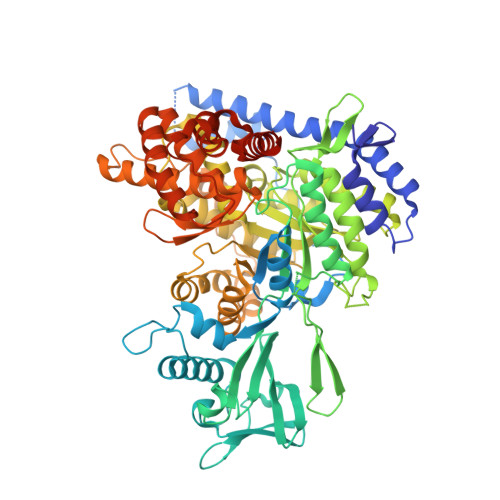
The glyoxylate shunt plays an important role in fatty acid metabolism and has been shown to be critical to survival of several pathogens involved in chronic infections. For Mycobacterium tuberculosis (Mtb), a strain with a defective glyoxylate shunt was previously shown to be unable to establish infection in a mouse model. We report the development of phenyl-diketo acid (PDKA) inhibitors of malate synthase (GlcB), one of two glyoxylate shunt enzymes, using structure-based methods. PDKA inhibitors were active against Mtb grown on acetate, and overexpression of GlcB ameliorated this inhibition. Crystal structures of complexes of GlcB with PDKA inhibitors guided optimization of potency. A selected PDKA compound demonstrated efficacy in a mouse model of tuberculosis. The discovery of these PDKA derivatives provides chemical validation of GlcB as an attractive target for tuberculosis therapeutics.
- Department of Biochemistry and Biophysics, Texas A&M University, College Station, TX 77843, USA.
Organizational Affiliation: