3J6P

Pseudo-atomic model of dynein microtubule binding domain-tubulin complex based on a cryoEM map


Experimental Data Snapshot

  • Method: ELECTRON MICROSCOPY
  • Resolution: 8.20 Å
  • Aggregation State: FILAMENT 
  • Reconstruction Method: HELICAL 

wwPDB Validation   3D Report Full Report


This is version 1.1 of the entry. See complete history


Literature

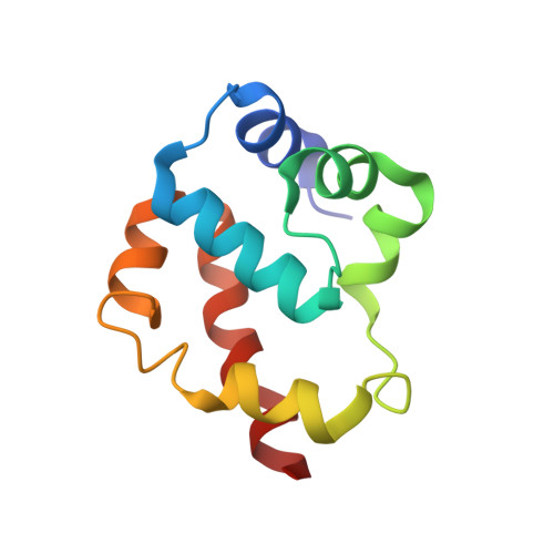
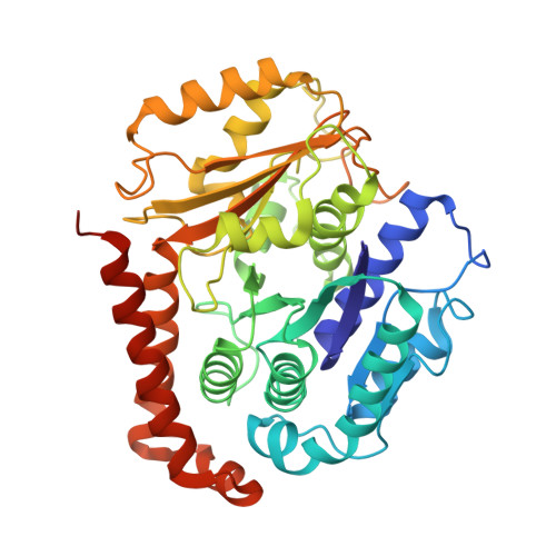
A flipped ion pair at the dynein-microtubule interface is critical for dynein motility and ATPase activation

Uchimura, S.Fujii, T.Takazaki, H.Ayukawa, R.Nishikawa, Y.Minoura, I.Hachikubo, Y.Kurisu, G.Sutoh, K.Kon, T.Namba, K.Muto, E.

(2015) J Cell Biol 208: 211-222

  • DOI: https://doi.org/10.1083/jcb.201407039
  • Primary Citation of Related Structures:  
    3J6P

  • PubMed Abstract: 
  • Dynein is a motor protein that moves on microtubules (MTs) using the energy of adenosine triphosphate (ATP) hydrolysis. To understand its motility mechanism, it is crucial to know how the signal of MT binding is transmitted to the ATPase domain to enhance ATP hydrolysis ...

    Dynein is a motor protein that moves on microtubules (MTs) using the energy of adenosine triphosphate (ATP) hydrolysis. To understand its motility mechanism, it is crucial to know how the signal of MT binding is transmitted to the ATPase domain to enhance ATP hydrolysis. However, the molecular basis of signal transmission at the dynein-MT interface remains unclear. Scanning mutagenesis of tubulin identified two residues in α-tubulin, R403 and E416, that are critical for ATPase activation and directional movement of dynein. Electron cryomicroscopy and biochemical analyses revealed that these residues form salt bridges with the residues in the dynein MT-binding domain (MTBD) that work in concert to induce registry change in the stalk coiled coil and activate the ATPase. The R403-E3390 salt bridge functions as a switch for this mechanism because of its reversed charge relative to other residues at the interface. This study unveils the structural basis for coupling between MT binding and ATPase activation and implicates the MTBD in the control of directional movement.


    Organizational Affiliation

    Laboratory for Molecular Biophysics, RIKEN Brain Science Institute, Wako, Saitama 351-0198, Japan emuto2@brain.riken.jp.


Macromolecules
Find similar proteins by:  (by identity cutoff)  |  3D Structure
Entity ID: 1
MoleculeChainsSequence LengthOrganismDetailsImage
Dynein heavy chain, cytoplasmicA [auth D]108Dictyostelium discoideumMutation(s): 0 
Gene Names: DDB_G0276355dhcA
UniProt
Find proteins for P34036 (Dictyostelium discoideum)
Explore P34036 
Go to UniProtKB:  P34036
Entity Groups  
Sequence Clusters30% Identity50% Identity70% Identity90% Identity95% Identity100% Identity
UniProt GroupP34036
Protein Feature View
Expand
  • Reference Sequence
Find similar proteins by:  (by identity cutoff)  |  3D Structure
Entity ID: 2
MoleculeChainsSequence LengthOrganismDetailsImage
Tubulin alpha-1A chainB [auth A]451Sus scrofaMutation(s): 0 
Gene Names: TUBA1A
UniProt
Find proteins for P02550 (Sus scrofa)
Explore P02550 
Go to UniProtKB:  P02550
Entity Groups  
Sequence Clusters30% Identity50% Identity70% Identity90% Identity95% Identity100% Identity
UniProt GroupP02550
Protein Feature View
Expand
  • Reference Sequence
Find similar proteins by:  (by identity cutoff)  |  3D Structure
Entity ID: 3
MoleculeChainsSequence LengthOrganismDetailsImage
Tubulin beta chainC [auth B]445Sus scrofaMutation(s): 0 
UniProt
Find proteins for P02554 (Sus scrofa)
Explore P02554 
Go to UniProtKB:  P02554
Entity Groups  
Sequence Clusters30% Identity50% Identity70% Identity90% Identity95% Identity100% Identity
UniProt GroupP02554
Protein Feature View
Expand
  • Reference Sequence
Small Molecules
Ligands 4 Unique
IDChainsName / Formula / InChI Key2D Diagram3D Interactions
TA1
Query on TA1

Download Ideal Coordinates CCD File 
G [auth B]TAXOL
C47 H51 N O14
RCINICONZNJXQF-MZXODVADSA-N
 Ligand Interaction
GTP
Query on GTP

Download Ideal Coordinates CCD File 
E [auth A]GUANOSINE-5'-TRIPHOSPHATE
C10 H16 N5 O14 P3
XKMLYUALXHKNFT-UUOKFMHZSA-N
 Ligand Interaction
GDP
Query on GDP

Download Ideal Coordinates CCD File 
F [auth B]GUANOSINE-5'-DIPHOSPHATE
C10 H15 N5 O11 P2
QGWNDRXFNXRZMB-UUOKFMHZSA-N
 Ligand Interaction
MG
Query on MG

Download Ideal Coordinates CCD File 
D [auth A]MAGNESIUM ION
Mg
JLVVSXFLKOJNIY-UHFFFAOYSA-N
 Ligand Interaction
Binding Affinity Annotations 
IDSourceBinding Affinity
TA1 BindingDB:  3J6P EC50: 520 (nM) from 1 assay(s)
Experimental Data & Validation

Experimental Data

  • Method: ELECTRON MICROSCOPY
  • Resolution: 8.20 Å
  • Aggregation State: FILAMENT 
  • Reconstruction Method: HELICAL 
EM Software:
TaskSoftware PackageVersion
RECONSTRUCTIONSPIDER

Structure Validation

View Full Validation Report


Entry History 

Deposition Data

Revision History  (Full details and data files)

  • Version 1.0: 2014-12-31
    Type: Initial release
  • Version 1.1: 2015-04-08
    Changes: Database references