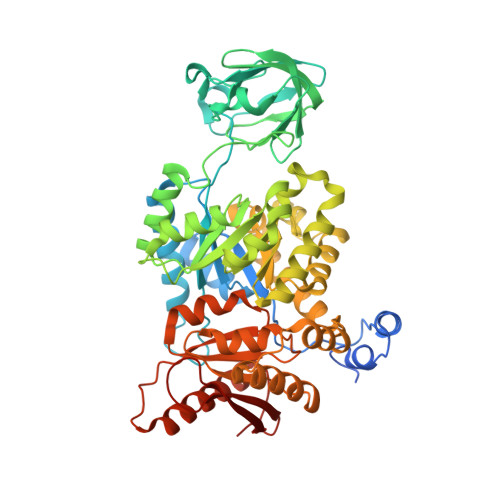
3GR4 | pdb_00003gr4

Activator-Bound Structure of Human Pyruvate Kinase M2


Experimental Data Snapshot

  • Method: X-RAY DIFFRACTION
  • Resolution: 1.60 Å
  • R-Value Free: 
    0.231 (Depositor), 0.235 (DCC) 
  • R-Value Work: 
    0.208 (Depositor), 0.212 (DCC) 
  • R-Value Observed: 
    0.208 (Depositor) 

Starting Model: experimental
View more details

wwPDB Validation 3D Report Full Report

Validation slider image for 3GR4

Ligand Structure Quality Assessment 


This is version 1.4 of the entry. See complete history

Literature

Macromolecule Content 

  • Total Structure Weight: 243.96 kDa 
  • Atom Count: 16,503 
  • Modeled Residue Count: 2,048 
  • Deposited Residue Count: 2,200 
  • Unique protein chains: 1

Macromolecules

Find similar proteins by:|  3D Structure
Entity ID: 1
MoleculeChains  Sequence LengthOrganismDetailsImage
Pyruvate kinase isozymes M1/M2
A, B, C, D
550Homo sapiensMutation(s): 0 
Gene Names: PKM2PK2PK3PKM
EC: 2.7.1.40 (PDB Primary Data), 2.7.11.1 (UniProt), 2.7.10.2 (UniProt)
UniProt & NIH Common Fund Data Resources
Find proteins for P14618 (Homo sapiens)
Explore P14618 
Go to UniProtKB:  P14618
PHAROS:  P14618
GTEx:  ENSG00000067225 
Entity Groups
Sequence Clusters30% Identity50% Identity70% Identity90% Identity95% Identity100% Identity
UniProt GroupP14618
Sequence Annotations
Expand
Reference Sequence

Small Molecules

Ligands 5 Unique
IDChains Name / Formula / InChI Key2D Diagram3D Interactions
DYY

Query on DYY


Download:Ideal Coordinates CCD File
CA [auth D],
G [auth A]
1-[(2,6-difluorophenyl)sulfonyl]-4-(2,3-dihydro-1,4-benzodioxin-6-ylsulfonyl)piperazine
C18 H18 F2 N2 O6 S2
SHWNKRPMUBFWKE-UHFFFAOYSA-N
ADP

Query on ADP


Download:Ideal Coordinates CCD File
N [auth A]ADENOSINE-5'-DIPHOSPHATE
C10 H15 N5 O10 P2
XTWYTFMLZFPYCI-KQYNXXCUSA-N
FBP

Query on FBP


Download:Ideal Coordinates CCD File
DA [auth D],
E [auth A],
O [auth B],
X [auth C]
1,6-di-O-phosphono-beta-D-fructofuranose
C6 H14 O12 P2
RNBGYGVWRKECFJ-ARQDHWQXSA-N
TLA

Query on TLA


Download:Ideal Coordinates CCD File
BA [auth C]
EA [auth D]
F [auth A]
JA [auth D]
P [auth B]
BA [auth C],
EA [auth D],
F [auth A],
JA [auth D],
P [auth B],
W [auth B],
Y [auth C]
L(+)-TARTARIC ACID
C4 H6 O6
FEWJPZIEWOKRBE-JCYAYHJZSA-N
UNX

Query on UNX


Download:Ideal Coordinates CCD File
AA [auth C]
FA [auth D]
GA [auth D]
H [auth A]
HA [auth D]
AA [auth C],
FA [auth D],
GA [auth D],
H [auth A],
HA [auth D],
I [auth A],
IA [auth D],
J [auth A],
K [auth A],
L [auth A],
M [auth A],
Q [auth B],
R [auth B],
S [auth B],
T [auth B],
U [auth B],
V [auth B],
Z [auth C]
UNKNOWN ATOM OR ION
X

Experimental Data & Validation

Experimental Data

  • Method: X-RAY DIFFRACTION
  • Resolution: 1.60 Å
  • R-Value Free:  0.231 (Depositor), 0.235 (DCC) 
  • R-Value Work:  0.208 (Depositor), 0.212 (DCC) 
  • R-Value Observed: 0.208 (Depositor) 
Space Group: P 1 21 1
Unit Cell:
Length ( Å )Angle ( ˚ )
a = 81.275α = 90
b = 153.206β = 102.91
c = 93.138γ = 90
Software Package:
Software NamePurpose
DENZOdata reduction
SCALEPACKdata scaling
REFMACrefinement
PDB_EXTRACTdata extraction
HKL-2000data reduction
HKL-2000data scaling
Cootmodel building
MolProbitymodel building
prodrgrefinement

Structure Validation

View Full Validation Report


Ligand Structure Quality Assessment 


Entry History 

Revision History  (Full details and data files)

  • Version 1.0: 2009-04-07
    Type: Initial release
  • Version 1.1: 2011-07-13
    Changes: Version format compliance
  • Version 1.2: 2017-11-01
    Changes: Refinement description
  • Version 1.3: 2020-07-29
    Type: Remediation
    Reason: Carbohydrate remediation
    Changes: Data collection, Database references, Derived calculations, Structure summary
  • Version 1.4: 2023-09-06
    Changes: Data collection, Database references, Refinement description, Structure summary