3AB3

Crystal structure of p115RhoGEF RGS domain in complex with G alpha 13


Experimental Data Snapshot

  • Method: X-RAY DIFFRACTION
  • Resolution: 2.40 Å
  • R-Value Free: 0.280 
  • R-Value Work: 0.205 
  • R-Value Observed: 0.205 

wwPDB Validation   3D Report Full Report


Ligand Structure Quality Assessment 


This is version 1.3 of the entry. See complete history


Literature

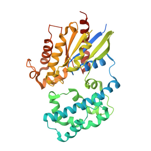
Identification of critical residues in G(alpha)13 for stimulation of p115RhoGEF activity and the structure of the G(alpha)13-p115RhoGEF regulator of G protein signaling homology (RH) domain complex.

Hajicek, N.Kukimoto-Niino, M.Mishima-Tsumagari, C.Chow, C.R.Shirouzu, M.Terada, T.Patel, M.Yokoyama, S.Kozasa, T.

(2011) J Biol Chem 286: 20625-20636

  • DOI: 10.1074/jbc.M110.201392
  • Primary Citation of Related Structures:  
    3AB3

  • PubMed Abstract: 
  • RH-RhoGEFs are a family of guanine nucleotide exchange factors that contain a regulator of G protein signaling homology (RH) domain. The heterotrimeric G protein Gα(13) stimulates the guanine nucleotide exchange factor (GEF) activity of RH-RhoGEFs, leading to activation of RhoA ...

    RH-RhoGEFs are a family of guanine nucleotide exchange factors that contain a regulator of G protein signaling homology (RH) domain. The heterotrimeric G protein Gα(13) stimulates the guanine nucleotide exchange factor (GEF) activity of RH-RhoGEFs, leading to activation of RhoA. The mechanism by which Gα(13) stimulates the GEF activity of RH-RhoGEFs, such as p115RhoGEF, has not yet been fully elucidated. Here, specific residues in Gα(13) that mediate activation of p115RhoGEF are identified. Mutation of these residues significantly impairs binding of Gα(13) to p115RhoGEF as well as stimulation of GEF activity. These data suggest that the exchange activity of p115RhoGEF is stimulated allosterically by Gα(13) and not through its interaction with a secondary binding site. A crystal structure of Gα(13) bound to the RH domain of p115RhoGEF is also presented, which differs from a previously crystallized complex with a Gα(13)-Gα(i1) chimera. Taken together, these data provide new insight into the mechanism by which p115RhoGEF is activated by Gα(13).


    Organizational Affiliation

    Department of Pharmacology, College of Medicine, University of Illinois, Chicago, Illinois 60612, USA.


Macromolecules
Find similar proteins by:  (by identity cutoff)  |  3D Structure
Entity ID: 1
MoleculeChainsSequence LengthOrganismDetailsImage
Guanine nucleotide-binding protein G(k) subunit alpha, Guanine nucleotide-binding protein subunit alpha-13A, C362Mus musculusMutation(s): 0 
Gene Names: Gna13Gna-13
UniProt & NIH Common Fund Data Resources
Find proteins for P27601 (Mus musculus)
Explore P27601 
Go to UniProtKB:  P27601
IMPC:  MGI:95768
Entity Groups  
Sequence Clusters30% Identity50% Identity70% Identity90% Identity95% Identity100% Identity
UniProt GroupP27601
Protein Feature View
Expand
  • Reference Sequence
Find similar proteins by:  (by identity cutoff)  |  3D Structure
Entity ID: 2
MoleculeChainsSequence LengthOrganismDetailsImage
Rho guanine nucleotide exchange factor 1B, D246Homo sapiensMutation(s): 0 
Gene Names: ARHGEF1
UniProt & NIH Common Fund Data Resources
Find proteins for Q92888 (Homo sapiens)
Explore Q92888 
Go to UniProtKB:  Q92888
PHAROS:  Q92888
Entity Groups  
Sequence Clusters30% Identity50% Identity70% Identity90% Identity95% Identity100% Identity
UniProt GroupQ92888
Protein Feature View
Expand
  • Reference Sequence
Small Molecules
Ligands 3 Unique
IDChainsName / Formula / InChI Key2D Diagram3D Interactions
GDP
Query on GDP

Download Ideal Coordinates CCD File 
F [auth A],
I [auth C]
GUANOSINE-5'-DIPHOSPHATE
C10 H15 N5 O11 P2
QGWNDRXFNXRZMB-UUOKFMHZSA-N
 Ligand Interaction
ALF
Query on ALF

Download Ideal Coordinates CCD File 
G [auth A],
J [auth C]
TETRAFLUOROALUMINATE ION
Al F4
UYOMQIYKOOHAMK-UHFFFAOYSA-J
 Ligand Interaction
MG
Query on MG

Download Ideal Coordinates CCD File 
E [auth A],
H [auth C]
MAGNESIUM ION
Mg
JLVVSXFLKOJNIY-UHFFFAOYSA-N
 Ligand Interaction
Experimental Data & Validation

Experimental Data

  • Method: X-RAY DIFFRACTION
  • Resolution: 2.40 Å
  • R-Value Free: 0.280 
  • R-Value Work: 0.205 
  • R-Value Observed: 0.205 
  • Space Group: P 1
Unit Cell:
Length ( Å )Angle ( ˚ )
a = 50.481α = 77.77
b = 70.64β = 84.45
c = 88.078γ = 80.11
Software Package:
Software NamePurpose
MOLREPphasing
CNSrefinement
HKL-2000data reduction
HKL-2000data scaling

Structure Validation

View Full Validation Report


Ligand Structure Quality Assessment 


Entry History 

Deposition Data

Revision History  (Full details and data files)

  • Version 1.0: 2010-12-29
    Type: Initial release
  • Version 1.1: 2011-07-13
    Changes: Version format compliance
  • Version 1.2: 2013-10-30
    Changes: Database references
  • Version 1.3: 2017-08-23
    Changes: Refinement description, Source and taxonomy