3TVJ | pdb_00003tvj

Catalytic fragment of MASP-2 in complex with its specific inhibitor developed by directed evolution on SGCI scaffold


Experimental Data Snapshot

  • Method: X-RAY DIFFRACTION
  • Resolution: 1.28 Å
  • R-Value Free: 
    0.205 (Depositor), 0.211 (DCC) 
  • R-Value Work: 
    0.156 (Depositor), 0.165 (DCC) 
  • R-Value Observed: 
    0.159 (Depositor) 

Starting Models: experimental
View more details

wwPDB Validation 3D Report Full Report

Validation slider image for 3TVJ

This is version 1.4 of the entry. See complete history

Literature

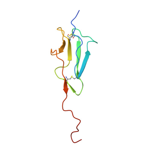
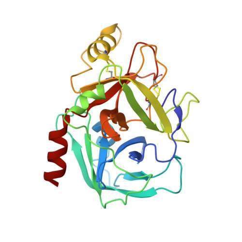
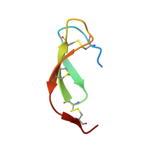
Monospecific Inhibitors Show That Both Mannan-binding Lectin-associated Serine Protease-1 (MASP-1) and -2 Are Essential for Lectin Pathway Activation and Reveal Structural Plasticity of MASP-2.

Heja, D.Harmat, V.Fodor, K.Wilmanns, M.Dobo, J.Kekesi, K.A.Zavodszky, P.Gal, P.Pal, G.

(2012) J Biological Chem 287: 20290-20300

  • DOI: https://doi.org/10.1074/jbc.M112.354332
  • Primary Citation Related Structures: 
    3TVJ, 4DJZ

  • PubMed Abstract: 

    The lectin pathway is an antibody-independent activation route of the complement system. It provides immediate defense against pathogens and altered self-cells, but it also causes severe tissue damage after stroke, heart attack, and other ischemia reperfusion injuries. The pathway is triggered by target binding of pattern recognition molecules leading to the activation of zymogen mannan-binding lectin-associated serine proteases (MASPs). MASP-2 is considered as the autonomous pathway-activator, while MASP-1 is considered as an auxiliary component. We evolved a pair of monospecific MASP inhibitors. In accordance with the key role of MASP-2, the MASP-2 inhibitor completely blocks the lectin pathway activation. Importantly, the MASP-1 inhibitor does the same, demonstrating that MASP-1 is not an auxiliary but an essential pathway component. We report the first Michaelis-like complex structures of MASP-1 and MASP-2 formed with substrate-like inhibitors. The 1.28 Å resolution MASP-2 structure reveals significant plasticity of the protease, suggesting that either an induced fit or a conformational selection mechanism should contribute to the extreme specificity of the enzyme.


  • Organizational Affiliation
    • Department of Biochemistry, Eötvös Loránd University, 1/C Pázmány Péter Street, H-1117, Budapest, Hungary.

Macromolecule Content 

  • Total Structure Weight: 39.88 kDa 
  • Atom Count: 3,450 
  • Modeled Residue Count: 360 
  • Deposited Residue Count: 366 
  • Unique protein chains: 3

Macromolecules

Find similar proteins by:|  3D Structure
Entity ID: 1
MoleculeChains  Sequence LengthOrganismDetailsImage
Mannan-binding lectin serine protease 2 A chain86Homo sapiensMutation(s): 0 
Gene Names: MASP2
EC: 3.4.21.104
UniProt & NIH Common Fund Data Resources
Find proteins for O00187 (Homo sapiens)
Explore O00187 
Go to UniProtKB:  O00187
PHAROS:  O00187
GTEx:  ENSG00000009724 
Entity Groups
Sequence Clusters30% Identity50% Identity70% Identity90% Identity95% Identity100% Identity
UniProt GroupO00187
Sequence Annotations
Expand
Reference Sequence
Find similar proteins by:|  3D Structure
Entity ID: 2
MoleculeChains  Sequence LengthOrganismDetailsImage
Mannan-binding lectin serine protease 2 B chain242Homo sapiensMutation(s): 0 
Gene Names: MASP2
EC: 3.4.21.104
UniProt & NIH Common Fund Data Resources
Find proteins for O00187 (Homo sapiens)
Explore O00187 
Go to UniProtKB:  O00187
PHAROS:  O00187
GTEx:  ENSG00000009724 
Entity Groups
Sequence Clusters30% Identity50% Identity70% Identity90% Identity95% Identity100% Identity
UniProt GroupO00187
Sequence Annotations
Expand
Reference Sequence
Find similar proteins by:|  3D Structure
Entity ID: 3
MoleculeChains  Sequence LengthOrganismDetailsImage
Protease inhibitor SGPI-2C [auth I]38Schistocerca gregariaMutation(s): 5 
Gene Names: GenBank: Y09605.1
UniProt
Find proteins for O46162 (Schistocerca gregaria)
Explore O46162 
Go to UniProtKB:  O46162
Entity Groups
Sequence Clusters30% Identity50% Identity70% Identity90% Identity95% Identity100% Identity
UniProt GroupO46162
Sequence Annotations
Expand
Reference Sequence

Small Molecules

Ligands 1 Unique
IDChains Name / Formula / InChI Key2D Diagram3D Interactions
SO4

Query on SO4


Download:Ideal Coordinates CCD File
D [auth B]SULFATE ION
O4 S
QAOWNCQODCNURD-UHFFFAOYSA-L

Experimental Data & Validation

Experimental Data

  • Method: X-RAY DIFFRACTION
  • Resolution: 1.28 Å
  • R-Value Free:  0.205 (Depositor), 0.211 (DCC) 
  • R-Value Work:  0.156 (Depositor), 0.165 (DCC) 
  • R-Value Observed: 0.159 (Depositor) 
Space Group: P 21 21 21
Unit Cell:
Length ( Å )Angle ( ˚ )
a = 50.916α = 90
b = 62.471β = 90
c = 114.189γ = 90
Software Package:
Software NamePurpose
MxCuBEdata collection
MOLREPphasing
REFMACrefinement
XDSdata reduction
XSCALEdata scaling

Structure Validation

View Full Validation Report


Entry History 

Deposition Data

Revision History  (Full details and data files)

  • Version 1.0: 2012-04-25
    Type: Initial release
  • Version 1.1: 2012-05-09
    Changes: Database references
  • Version 1.2: 2012-06-27
    Changes: Database references
  • Version 1.3: 2023-09-13
    Changes: Data collection, Database references, Derived calculations, Refinement description
  • Version 1.4: 2024-11-20
    Changes: Structure summary