3TI1 | pdb_00003ti1

CDK2 in complex with SUNITINIB


Experimental Data Snapshot

  • Method: X-RAY DIFFRACTION
  • Resolution: 1.99 Å
  • R-Value Free: 
    0.262 (Depositor), 0.256 (DCC) 
  • R-Value Work: 
    0.205 (Depositor), 0.200 (DCC) 

Starting Model: experimental
View more details

wwPDB Validation 3D Report Full Report

Validation slider image for 3TI1

Ligand Structure Quality Assessment 


This is version 1.3 of the entry. See complete history

Literature

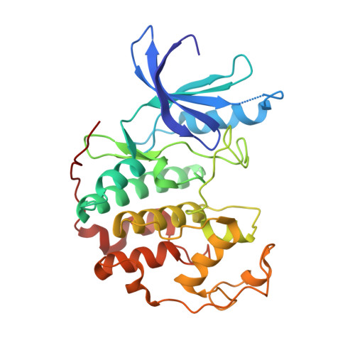
A Novel Approach to the Discovery of Small-Molecule Ligands of CDK2.

Martin, M.P.Alam, R.Betzi, S.Ingles, D.J.Zhu, J.Y.Schonbrunn, E.

(2012) Chembiochem 13: 2128-2136

  • DOI: https://doi.org/10.1002/cbic.201200316
  • Primary Citation Related Structures: 
    3TI1, 3TIY, 3TIZ, 4ERW, 4EZ3, 4EZ7

  • PubMed Abstract: 

    In an attempt to identify novel small-molecule ligands of cyclin-dependent kinase 2 (CDK2) with potential as allosteric inhibitors, we have devised a robust and cost-effective fluorescence-based high-throughput screening assay. The assay is based on the specific interaction of CDK2 with the extrinsic fluorophore 8-anilino-1-naphthalene sulfonate (ANS), which binds to a large allosteric pocket adjacent to the ATP site. Hit compounds that displace ANS directly or indirectly from CDK2 are readily classified as ATP site binders or allosteric ligands through the use of staurosporine, which blocks the ATP site without displacing ANS. Pilot screening of 1453 compounds led to the discovery of 12 compounds with displacement activities (EC(50) values) ranging from 6 to 44 μM, all of which were classified as ATP-site-directed ligands. Four new type I inhibitor scaffolds were confirmed by X-ray crystallography. Although this small compound library contained only ATP-site-directed ligands, the application of this assay to large compound libraries has the potential to reveal previously unrecognized chemical scaffolds suitable for structure-based design of CDK2 inhibitors with new mechanisms of action.


  • Organizational Affiliation
    • Drug Discovery Department, Moffitt Cancer Center and Research Institute, 12902 Magnolia Drive, Tampa, FL 33612, USA.

Macromolecule Content 

  • Total Structure Weight: 34.87 kDa 
  • Atom Count: 2,528 
  • Modeled Residue Count: 294 
  • Deposited Residue Count: 298 
  • Unique protein chains: 1

Macromolecules

Find similar proteins by:|  3D Structure
Entity ID: 1
MoleculeChains  Sequence LengthOrganismDetailsImage
Cyclin-dependent kinase 2298Homo sapiensMutation(s): 0 
Gene Names: CDK2
EC: 2.7.11.22
UniProt & NIH Common Fund Data Resources
Find proteins for P24941 (Homo sapiens)
Explore P24941 
Go to UniProtKB:  P24941
PHAROS:  P24941
GTEx:  ENSG00000123374 
Entity Groups
Sequence Clusters30% Identity50% Identity70% Identity90% Identity95% Identity100% Identity
UniProt GroupP24941
Sequence Annotations
Expand
Reference Sequence

Small Molecules

Ligands 2 Unique
IDChains Name / Formula / InChI Key2D Diagram3D Interactions
B49

Query on B49


Download:Ideal Coordinates CCD File
B [auth A]N-[2-(diethylamino)ethyl]-5-[(Z)-(5-fluoro-2-oxo-1,2-dihydro-3H-indol-3-ylidene)methyl]-2,4-dimethyl-1H-pyrrole-3-carbo xamide
C22 H27 F N4 O2
WINHZLLDWRZWRT-ATVHPVEESA-N
EDO

Query on EDO


Download:Ideal Coordinates CCD File
C [auth A]
D [auth A]
E [auth A]
F [auth A]
G [auth A]
C [auth A],
D [auth A],
E [auth A],
F [auth A],
G [auth A],
H [auth A],
I [auth A],
J [auth A]
1,2-ETHANEDIOL
C2 H6 O2
LYCAIKOWRPUZTN-UHFFFAOYSA-N

Experimental Data & Validation

Experimental Data

  • Method: X-RAY DIFFRACTION
  • Resolution: 1.99 Å
  • R-Value Free:  0.262 (Depositor), 0.256 (DCC) 
  • R-Value Work:  0.205 (Depositor), 0.200 (DCC) 
Space Group: P 21 21 21
Unit Cell:
Length ( Å )Angle ( ˚ )
a = 53.56α = 90
b = 71.94β = 90
c = 72.46γ = 90
Software Package:
Software NamePurpose
PHENIXmodel building
PHENIXrefinement
XDSdata reduction
XDSdata scaling
PHENIXphasing

Structure Validation

View Full Validation Report


Ligand Structure Quality Assessment 


Entry History 

Deposition Data

Revision History  (Full details and data files)

  • Version 1.0: 2012-08-22
    Type: Initial release
  • Version 1.1: 2012-08-29
    Changes: Database references
  • Version 1.2: 2012-10-10
    Changes: Database references
  • Version 1.3: 2023-09-13
    Changes: Data collection, Database references, Derived calculations, Refinement description, Structure summary