3RWA

Crystal structure of circular-permutated mKate


Experimental Data Snapshot

  • Method: X-RAY DIFFRACTION
  • Resolution: 1.67 Å
  • R-Value Free: 0.215 
  • R-Value Work: 0.182 
  • R-Value Observed: 0.182 

wwPDB Validation   3D Report Full Report


This is version 1.6 of the entry. See complete history


Literature

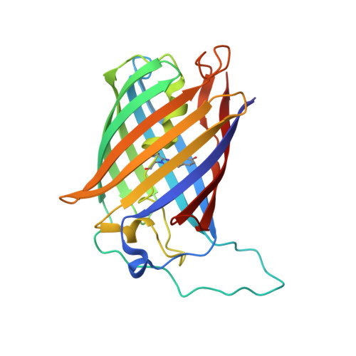
Circular permutation of red fluorescent proteins.

Shui, B.Wang, Q.Lee, F.Byrnes, L.J.Chudakov, D.M.Lukyanov, S.A.Sondermann, H.Kotlikoff, M.I.

(2011) PLoS One 6: e20505-e20505

  • DOI: https://doi.org/10.1371/journal.pone.0020505
  • Primary Citation of Related Structures:  
    3RWA, 3RWT

  • PubMed Abstract: 

    Circular permutation of fluorescent proteins provides a substrate for the design of molecular sensors. Here we describe a systematic exploration of permutation sites for mCherry and mKate using a tandem fusion template approach. Circular permutants retaining more than 60% (mCherry) and 90% (mKate) brightness of the parent molecules are reported, as well as a quantitative evaluation of the fluorescence from neighboring mutations. Truncations of circular permutants indicated essential N- and C-terminal segments and substantial flexibility in the use of these molecules. Structural evaluation of two cp-mKate variants indicated no major conformational changes from the previously reported wild-type structure, and cis conformation of the chromophores. Four cp-mKates were identified with over 80% of native fluorescence, providing important new building blocks for sensor and complementation experiments.


  • Organizational Affiliation

    Department of Biomedical Sciences, College of Veterinary Medicine, Cornell University, Ithaca, New York, United States of America.


Macromolecules
Find similar proteins by:  (by identity cutoff)  |  3D Structure
Entity ID: 1
MoleculeChains Sequence LengthOrganismDetailsImage
Fluorescent protein FP480
A, B, C, D, E
A, B, C, D, E, F, G, H
233Entacmaea quadricolorMutation(s): 1 
UniProt
Find proteins for D0VX33 (Entacmaea quadricolor)
Explore D0VX33 
Go to UniProtKB:  D0VX33
Entity Groups  
Sequence Clusters30% Identity50% Identity70% Identity90% Identity95% Identity100% Identity
UniProt GroupD0VX33
Sequence Annotations
Expand
  • Reference Sequence
Small Molecules
Modified Residues  1 Unique
IDChains TypeFormula2D DiagramParent
NRQ
Query on NRQ
A, B, C, D, E
A, B, C, D, E, F, G, H
L-PEPTIDE LINKINGC16 H17 N3 O4 SMET, TYR, GLY
Experimental Data & Validation

Experimental Data

  • Method: X-RAY DIFFRACTION
  • Resolution: 1.67 Å
  • R-Value Free: 0.215 
  • R-Value Work: 0.182 
  • R-Value Observed: 0.182 
  • Space Group: C 1 2 1
Unit Cell:
Length ( Å )Angle ( ˚ )
a = 109.357α = 90
b = 93.28β = 96.52
c = 194.37γ = 90
Software Package:
Software NamePurpose
PHENIXrefinement
SCALEPACKdata scaling
PHASERphasing
REFMACrefinement
PDB_EXTRACTdata extraction
DENZOdata reduction

Structure Validation

View Full Validation Report


Entry History 

Deposition Data

Revision History  (Full details and data files)

  • Version 1.0: 2011-06-15
    Type: Initial release
  • Version 1.1: 2011-07-13
    Changes: Version format compliance
  • Version 1.2: 2011-09-14
    Changes: Database references
  • Version 1.3: 2011-11-16
    Changes: Atomic model
  • Version 1.4: 2017-06-21
    Changes: Advisory, Database references, Derived calculations, Source and taxonomy
  • Version 1.5: 2017-11-08
    Changes: Advisory, Refinement description
  • Version 1.6: 2019-07-17
    Changes: Data collection, Derived calculations, Refinement description