3RB8

Structure of the phage tubulin PhuZ(SeMet)-GDP


Experimental Data Snapshot

  • Method: X-RAY DIFFRACTION
  • Resolution: 2.60 Å
  • R-Value Free: 0.237 
  • R-Value Work: 0.163 
  • R-Value Observed: 0.171 

wwPDB Validation   3D Report Full Report


Ligand Structure Quality Assessment 


This is version 1.1 of the entry. See complete history


Literature

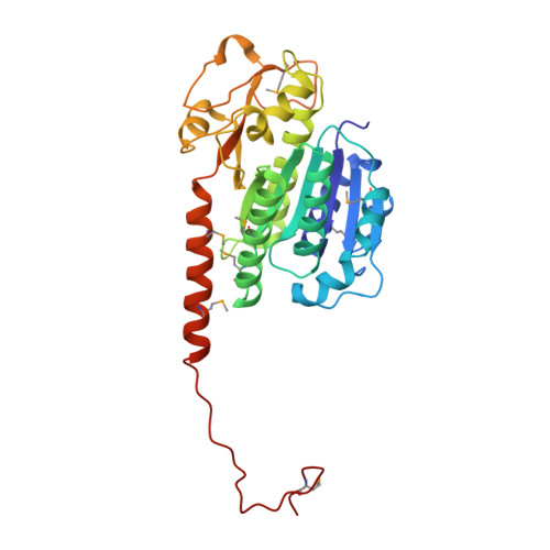
A phage tubulin assembles dynamic filaments by an atypical mechanism to center viral DNA within the host cell.

Kraemer, J.A.Erb, M.L.Waddling, C.A.Montabana, E.A.Zehr, E.A.Wang, H.Nguyen, K.Pham, D.S.Agard, D.A.Pogliano, J.

(2012) Cell 149: 1488-1499

  • DOI: https://doi.org/10.1016/j.cell.2012.04.034
  • Primary Citation of Related Structures:  
    3R4V, 3RB8

  • PubMed Abstract: 

    Tubulins are essential for the reproduction of many eukaryotic viruses, but historically, bacteriophage were assumed not to require a cytoskeleton. Here, we identify a tubulin-like protein, PhuZ, from bacteriophage 201φ2-1 and show that it forms filaments in vivo and in vitro. The PhuZ structure has a conserved tubulin fold, with an unusual, extended C terminus that we demonstrate to be critical for polymerization in vitro and in vivo. Longitudinal packing in the crystal lattice mimics packing observed by EM of in-vitro-formed filaments, indicating how interactions between the C terminus and the following monomer drive polymerization. PhuZ forms a filamentous array that is required for positioning phage DNA within the bacterial cell. Correct positioning to the cell center and optimal phage reproduction only occur when the PhuZ filament is dynamic. Thus, we show that PhuZ assembles a spindle-like array that functions analogously to the microtubule-based spindles of eukaryotes.


  • Organizational Affiliation

    Department of Biochemistry and Biophysics and the Howard Hughes Medical Institute, University of California, San Francisco, San Francisco, CA 94158, USA.


Macromolecules
Find similar proteins by:  (by identity cutoff)  |  3D Structure
Entity ID: 1
MoleculeChains Sequence LengthOrganismDetailsImage
Putative uncharacterized protein315Pseudomonas phage 201phi2-1Mutation(s): 0 
Gene Names: 201phi2-1p059
UniProt
Find proteins for B3FK34 (Pseudomonas phage 201phi2-1)
Explore B3FK34 
Go to UniProtKB:  B3FK34
Entity Groups  
Sequence Clusters30% Identity50% Identity70% Identity90% Identity95% Identity100% Identity
UniProt GroupB3FK34
Sequence Annotations
Expand
  • Reference Sequence
Small Molecules
Ligands 2 Unique
IDChains Name / Formula / InChI Key2D Diagram3D Interactions
GDP
Query on GDP

Download Ideal Coordinates CCD File 
B [auth A]GUANOSINE-5'-DIPHOSPHATE
C10 H15 N5 O11 P2
QGWNDRXFNXRZMB-UUOKFMHZSA-N
MG
Query on MG

Download Ideal Coordinates CCD File 
C [auth A]MAGNESIUM ION
Mg
JLVVSXFLKOJNIY-UHFFFAOYSA-N
Modified Residues  1 Unique
IDChains TypeFormula2D DiagramParent
MSE
Query on MSE
A
L-PEPTIDE LINKINGC5 H11 N O2 SeMET
Experimental Data & Validation

Experimental Data

  • Method: X-RAY DIFFRACTION
  • Resolution: 2.60 Å
  • R-Value Free: 0.237 
  • R-Value Work: 0.163 
  • R-Value Observed: 0.171 
  • Space Group: P 21 21 21
Unit Cell:
Length ( Å )Angle ( ˚ )
a = 46.981α = 90
b = 76.062β = 90
c = 92.758γ = 90
Software Package:
Software NamePurpose
Blu-Icedata collection
SOLVEphasing
PHENIXrefinement
MOSFLMdata reduction
SCALAdata scaling

Structure Validation

View Full Validation Report


Ligand Structure Quality Assessment 


Entry History 

Deposition Data

Revision History  (Full details and data files)

  • Version 1.0: 2012-07-04
    Type: Initial release
  • Version 1.1: 2013-01-23
    Changes: Database references