Structural and biochemical insights into the mechanism of fosfomycin phosphorylation by fosfomycin resistance kinase FomA.
Pakhomova, S., Bartlett, S.G., Doerner, P.A., Newcomer, M.E.(2011) Biochemistry 50: 6909-6919
- PubMed: 21728358 Search on PubMedSearch on PubMed Central
- DOI: https://doi.org/10.1021/bi2004334
- Primary Citation Related Structures:
3QUN, 3QUO, 3QUR, 3QVF, 3QVH - PubMed Abstract:
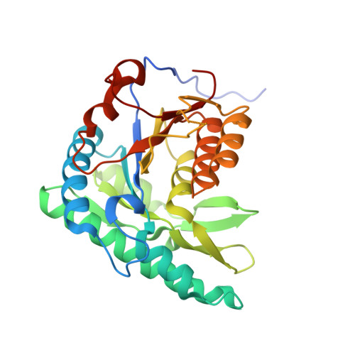
We present here the crystal structures of fosfomycin resistance protein (FomA) complexed with MgATP, with ATP and fosfomycin, with MgADP and fosfomycin vanadate, with MgADP and the product of the enzymatic reaction, fosfomycin monophosphate, and with ADP at 1.87, 1.58, 1.85, 1.57, and 1.85 Å resolution, respectively. Structures of these complexes that approximate different reaction steps allowed us to distinguish the catalytically active conformation of ATP and to reconstruct the model of the MgATP·fosfomycin complex. According to the model, the triphosphate tail of the nucleotide is aligned toward the phosphonate moiety of fosfomycin, in contest to the previously published MgAMPPNP complex, with the attacking fosfomycin oxygen positioned 4 Å from the γ-phosphorus of ATP. Site-directed mutagenesis studies and comparison of these structures with that of homologous N-acetyl-l-glutamate and isopentenyl phosphate kinases allowed us to propose a model of phosphorylation of fosfomycin by FomA enzyme. A Mg cation ligates all three phosphate groups of ATP and together with positively charged K216, K9, K18, and H58 participates in the dissipation of negative charge during phosphoryl transfer, indicating that the transferred phosphate group is highly negatively charged, which would be expected for an associative mechanism. K216 polarizes the γ-phosphoryl group of ATP. K9, K18, and H58 participate in stabilization of the transition state. D150 and D208 play organizational roles in catalysis. S148, S149, and T210 participate in fosfomycin binding, with T210 being crucial for catalysis. Hence, it appears that as in the homologous enzymes, FomA-catalyzed phosphoryl transfer takes place by an in-line predominantly associative mechanism.
- Department of Biological Sciences, Louisiana State University, Baton Rouge, Louisiana 70803, United States. sveta@lsu.edu
Organizational Affiliation: