3PLF

Reverse Binding Mode of MetRD peptide complexed with c-Cbl TKB domain


Experimental Data Snapshot

  • Method: X-RAY DIFFRACTION
  • Resolution: 1.92 Å
  • R-Value Free: 0.180 
  • R-Value Work: 0.159 
  • R-Value Observed: 0.160 

wwPDB Validation   3D Report Full Report


This is version 1.5 of the entry. See complete history


Literature

An adjacent arginine, and the phosphorylated tyrosine in the c-Met receptor target sequence, dictates the orientation of c-Cbl binding

Sun, Q.Ng, C.Guy, G.R.Sivaraman, J.

(2011) FEBS Lett 585: 281-285

  • DOI: https://doi.org/10.1016/j.febslet.2010.11.060
  • Primary Citation of Related Structures:  
    3PLF

  • PubMed Abstract: 

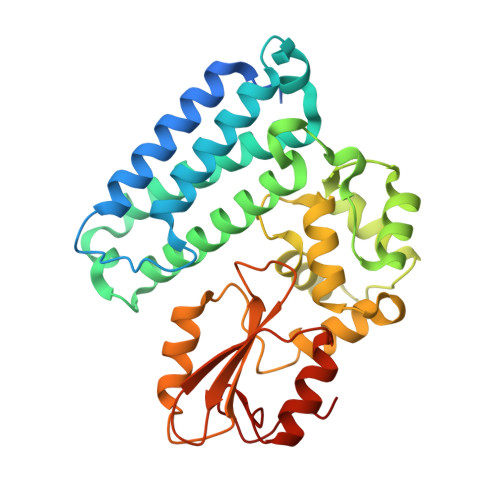
    Previously, we have demonstrated that the tyrosine phosphorylated hepatocyte growth factor receptor (Met) binds to the c-Cbl phosphotyrosine-recognition, tyrosine kinase binding (TKB) domain in a reverse orientation compared to other c-Cbl binding partners. A Met peptide with the DpYR motif changed to RpYD (MetRD) retains a similar TKB binding affinity as the native Met peptide. However, the TKB: MetRD complex crystal structure reveals a complete reversal of the binding orientation. Collated data indicates that both binding and orientation is dictated by the phosphorylated tyrosine and an adjacent arginine forming intra-peptide hydrogen bonds and aligning unidirectionally with complementary charges in the phosphotyrosine binding pocket of c-Cbl.


  • Organizational Affiliation

    Department of Biological Sciences, National University of Singapore, Singapore, Singapore.


Macromolecules

Find similar proteins by:  Sequence   |   3D Structure  

Entity ID: 1
MoleculeChains Sequence LengthOrganismDetailsImage
MetRD peptide
A, C
11N/AMutation(s): 0 
Entity Groups  
Sequence Clusters30% Identity50% Identity70% Identity90% Identity95% Identity100% Identity
Sequence Annotations
Expand
  • Reference Sequence
Find similar proteins by:  (by identity cutoff)  |  3D Structure
Entity ID: 2
MoleculeChains Sequence LengthOrganismDetailsImage
E3 ubiquitin-protein ligase CBL
B, D
329Homo sapiensMutation(s): 0 
Gene Names: c-Cbl
EC: 6.3.2
UniProt & NIH Common Fund Data Resources
Find proteins for P22681 (Homo sapiens)
Explore P22681 
Go to UniProtKB:  P22681
PHAROS:  P22681
GTEx:  ENSG00000110395 
Entity Groups  
Sequence Clusters30% Identity50% Identity70% Identity90% Identity95% Identity100% Identity
UniProt GroupP22681
Sequence Annotations
Expand
  • Reference Sequence
Experimental Data & Validation

Experimental Data

  • Method: X-RAY DIFFRACTION
  • Resolution: 1.92 Å
  • R-Value Free: 0.180 
  • R-Value Work: 0.159 
  • R-Value Observed: 0.160 
  • Space Group: P 1 21 1
Unit Cell:
Length ( Å )Angle ( ˚ )
a = 52.743α = 90
b = 104.483β = 90.19
c = 60.548γ = 90
Software Package:
Software NamePurpose
DENZOdata reduction
SCALEPACKdata scaling
MOLREPphasing
REFMACrefinement
PDB_EXTRACTdata extraction
HKL-2000data collection
HKL-2000data reduction
HKL-2000data scaling

Structure Validation

View Full Validation Report


Entry History 

Deposition Data

Revision History  (Full details and data files)

  • Version 1.0: 2010-12-01
    Type: Initial release
  • Version 1.1: 2011-07-13
    Changes: Version format compliance
  • Version 1.2: 2013-06-05
    Changes: Database references
  • Version 1.3: 2017-10-04
    Changes: Data collection, Refinement description
  • Version 1.4: 2023-11-01
    Changes: Data collection, Database references, Derived calculations, Refinement description
  • Version 1.5: 2023-12-06
    Changes: Data collection