3OKA

Crystal structure of Corynebacterium glutamicum PimB' in complex with GDP-Man (triclinic crystal form)


Experimental Data Snapshot

  • Method: X-RAY DIFFRACTION
  • Resolution: 2.20 Å
  • R-Value Free: 0.221 
  • R-Value Work: 0.189 
  • R-Value Observed: 0.191 

wwPDB Validation   3D Report Full Report


Ligand Structure Quality Assessment 


This is version 1.2 of the entry. See complete history


Literature

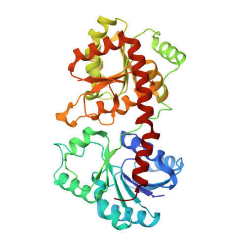
Acceptor substrate discrimination in phosphatidyl-myo-inositol mannoside synthesis: structural and mutational analysis of mannosyltransferase Corynebacterium glutamicum PimB'.

Batt, S.M.Jabeen, T.Mishra, A.K.Veerapen, N.Krumbach, K.Eggeling, L.Besra, G.S.Futterer, K.

(2010) J Biol Chem 285: 37741-37752

  • DOI: https://doi.org/10.1074/jbc.M110.165407
  • Primary Citation of Related Structures:  
    3OKA, 3OKC, 3OKP

  • PubMed Abstract: 

    Long term survival of the pathogen Mycobacterium tuberculosis in humans is linked to the immunomodulatory potential of its complex cell wall glycolipids, which include the phosphatidylinositol mannoside (PIM) series as well as the related lipomannan and lipoarabinomannan glycoconjugates. PIM biosynthesis is initiated by a set of cytosolic α-mannosyltransferases, catalyzing glycosyl transfer from the activated saccharide donor GDP-α-D-mannopyranose to the acceptor phosphatidyl-myo-inositol (PI) in an ordered and regio-specific fashion. Herein, we report the crystal structure of mannosyltransferase Corynebacterium glutamicum PimB' in complex with nucleotide to a resolution of 2.0 Å. PimB' attaches mannosyl selectively to the 6-OH of the inositol moiety of PI. Two crystal forms and GDP- versus GDP-α-d-mannopyranose-bound complexes reveal flexibility of the nucleotide conformation as well as of the structural framework of the active site. Structural comparison, docking of the saccharide acceptor, and site-directed mutagenesis pin regio-selectivity to a conserved Asp residue in the N-terminal domain that forces presentation of the correct inositol hydroxyl to the saccharide donor.


  • Organizational Affiliation

    School of Biosciences, University of Birmingham, Edgbaston, Birmingham B15 2TT, United Kingdom.


Macromolecules
Find similar proteins by:  (by identity cutoff)  |  3D Structure
Entity ID: 1
MoleculeChains Sequence LengthOrganismDetailsImage
GDP-mannose-dependent alpha-(1-6)-phosphatidylinositol monomannoside mannosyltransferase
A, B
381Corynebacterium glutamicumMutation(s): 0 
Gene Names: pimBCgl2186cg2400
EC: 2.4.1.57
UniProt
Find proteins for Q8NNK8 (Corynebacterium glutamicum (strain ATCC 13032 / DSM 20300 / BCRC 11384 / JCM 1318 / LMG 3730 / NCIMB 10025))
Explore Q8NNK8 
Go to UniProtKB:  Q8NNK8
Entity Groups  
Sequence Clusters30% Identity50% Identity70% Identity90% Identity95% Identity100% Identity
UniProt GroupQ8NNK8
Sequence Annotations
Expand
  • Reference Sequence

Find similar proteins by:  Sequence   |   3D Structure  

Entity ID: 2
MoleculeChains Sequence LengthOrganismDetailsImage
N-terminal His-affinity tag
C, D
21Escherichia coliMutation(s): 0 
Gene Names: His-affinity tag
Entity Groups  
Sequence Clusters30% Identity50% Identity70% Identity90% Identity95% Identity100% Identity
Sequence Annotations
Expand
  • Reference Sequence
Small Molecules
Ligands 3 Unique
IDChains Name / Formula / InChI Key2D Diagram3D Interactions
GDD
Query on GDD

Download Ideal Coordinates CCD File 
H [auth A],
N [auth B]
GUANOSINE-5'-DIPHOSPHATE-ALPHA-D-MANNOSE
C16 H25 N5 O16 P2
MVMSCBBUIHUTGJ-GDJBGNAASA-N
SO4
Query on SO4

Download Ideal Coordinates CCD File 
E [auth A]
F [auth A]
G [auth A]
J [auth B]
K [auth B]
E [auth A],
F [auth A],
G [auth A],
J [auth B],
K [auth B],
L [auth B],
M [auth B],
O [auth C],
P [auth D],
Q [auth D]
SULFATE ION
O4 S
QAOWNCQODCNURD-UHFFFAOYSA-L
GOL
Query on GOL

Download Ideal Coordinates CCD File 
I [auth A]GLYCEROL
C3 H8 O3
PEDCQBHIVMGVHV-UHFFFAOYSA-N
Binding Affinity Annotations 
IDSourceBinding Affinity
GDD PDBBind:  3OKA Kd: 1.90e+4 (nM) from 1 assay(s)
Binding MOAD:  3OKA Kd: 1.90e+4 (nM) from 1 assay(s)
Experimental Data & Validation

Experimental Data

  • Method: X-RAY DIFFRACTION
  • Resolution: 2.20 Å
  • R-Value Free: 0.221 
  • R-Value Work: 0.189 
  • R-Value Observed: 0.191 
  • Space Group: P 1
Unit Cell:
Length ( Å )Angle ( ˚ )
a = 44.07α = 92.05
b = 50.02β = 92.68
c = 85.3γ = 89.94
Software Package:
Software NamePurpose
ADSCdata collection
SHARPphasing
REFMACrefinement
XDSdata reduction
XSCALEdata scaling

Structure Validation

View Full Validation Report


Ligand Structure Quality Assessment 


Entry History 

Deposition Data

Revision History  (Full details and data files)

  • Version 1.0: 2010-09-15
    Type: Initial release
  • Version 1.1: 2011-07-13
    Changes: Version format compliance
  • Version 1.2: 2024-02-21
    Changes: Data collection, Database references, Derived calculations